
- Title
-
Gata4 regulates the formation of multiple organs
- Authors
- Holtzinger, A., and Evans, T.
- Source
- Full text @ Development
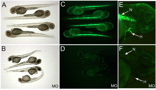
The Gata4 morpholino effectively blocks GFP translation in the gata4:GFP line, and generates a consistent phenotype. (A,C,E) Uninjected control fish; (B,D,F) embryos derived from eggs injected with MO. Living embryos are shown at 2 dpf in brightfield (A,B) or under fluorescence (C-F). (E,F) Control (E) and morphant (F) embryos at 3 dpf (lateral views, dorsal to the top with anterior to the right), demonstrating the complete block to GFP expression in both the notochord (N) and the heart (H).
|
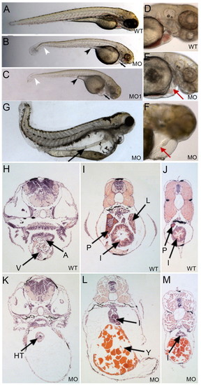
Gata4 deficient embryos develop cardiac and gut tubes but no derivative organs. (A-C) Shown at 3 dpf are (A) a representative control embryo (WT), and embryos derived from eggs injected with (B) the morpholino targeting the Gata4 5'UTR (MO) or (C) the morpholino targeting an intron/exon boundary (MO1). The morphant fish display a kink in the tail (white arrowheads), a pericardial edema (black arrows), and regression of the yolk stalk extension (black arrowheads). (D,E) Control embryo (D) and a representative morphant (E), showing a relative lack of blood in the linear but constricted heart tube at 3 dpf in the morphant (red arrow). (F) At 5 dpf, the morphant linear heart tube (red arrow) is distended. Views are lateral, dorsal to the top and anterior to the right. (G) At 8 dpf, a linear gut tube is shown (arrow), running the length of the morphant body. (H-M) Representative histological cross sections through control (WT) or morphant (MO) embryos. Equivalent sections are progressively more caudal, at the level of the heart (H,K), mid-pancreas (I,L) or the caudal end of the swim bladder (J,M). A, atrium; V, ventricle; HT, heart tube; P, pancreas; L, liver; I, intestine; Y, yolk. |
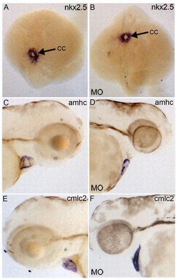
Heart tube formation is normal, but morphogenesis fails in Gata4 deficient embryos. Control uninjected embryos (A,C,E) are compared with embryos derived from eggs injected with MO (B,D,F). Embryos were processed for in situ hybridization to detect transcripts for Nkx2.5 (A,B), Amhc (C,D) or Cmlc2 (E,F). The morphant embryos show normal formation of the cardiac cone (cc) at the 20-somite stage (B). Likewise, the cardiomyocyte markers Amhc and Cmlc2 are expressed normally, shown here at 3 dpf. (A,B) Dorsal views with anterior to the top; (C,D) lateral views with anterior to the right and dorsal to the top; (E,F) lateral views with anterior to the left and dorsal to the top. Embryos are shown in both orientations to show the lack of looping in the linear heart tube of the morphant embryos. EXPRESSION / LABELING:
|
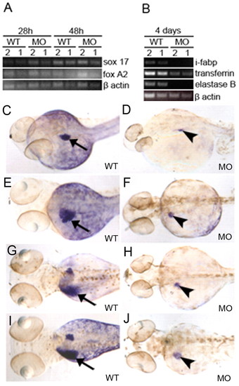
Gata4 deficient embryos express early endoderm markers but organogenesis fails. (A) Shown are representative semi-quantitative RT-PCR results for the early endoderm markers Sox17 and FoxA2. Assays used either 2 or 1 µg of RNA, isolated for the RT reaction at 28 hpf or 48 hpf, as indicated. We consistently found no significant difference in the expression levels comparing control (WT) and morphant (MO) embryos. (B) RT-PCR assays were also performed using RNA isolated at 4 dpf for markers of differentiated intestine (I-fabp), liver (transferrin), and pancreas (elastaseB). (C-J) Representative embryos are shown following in situ hybridization to detect transferrin RNA, comparing control (WT) and morphant (MO) embryos at 2 dpf (C,D), 3 dpf (E,F), 4 dpf (G,H) and 5 dpf (I,J). In all cases, the morphant embryos develop with a small patch of transferrin-positive cells (arrowheads in MO panels), but this patch fails to grow when compared with controls (arrows in WT panels). Views are dorsal, anterior to the left. |
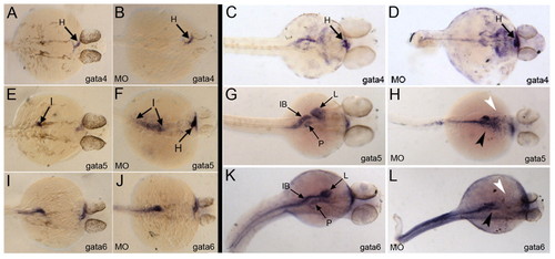
Gata4 deficient fish are blocked at a relatively late stage of gut tube morphogenesis. Shown are representative examples of control embryos (A,C,E,G,I,K) and corresponding morphant (MO) embryos (B,D,F,H,J,L) following in situ hybridization using probes to detect Gata4 (A-D), Gata5 (E-H) or Gata6 (I-L). Embryos were fixed at 28 hpf (A,B,E,F,I,J) or 48 hpf (C,D,G,H,K,L). Note that, at 28 hpf, Gata5 expression is enhanced in the morphant embryos (arrows in F), whereas at 48 hpf Gata4 transcript levels are clearly increased (D), but Gata5 and Gata6 expression (H,L) is missing in the regions that normally show signal for pancreas (black arrowhead) and liver (white arrowhead). Views are dorsal, with anterior to the right. I, intestinal rod; H, heart; IB, intestinal bulb; L, liver; P, pancreas. |
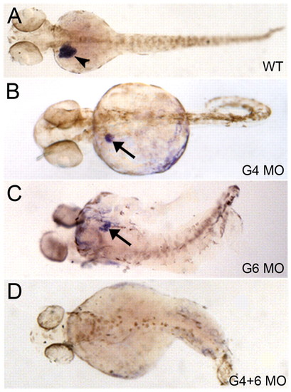
Gata4 and Gata6 have redundant functions for liver development, but each gene is essential for liver bud growth. Representative control embryos (A, WT), and embryos injected with the morpholinos specific for Gata4 (B, G4 MO), Gata6 (C, G6 MO), or both (D, G4 +6 MO), are shown following in situ hybridization to detect transcript for the liver differentiation marker transferrin. At 3 dpf, the control embryos show a much enhanced liver growth (arrowhead) compared with either the Gata4 or Gata6 morphants (arrows). However, both morphants consistently show small liver buds. By contrast, injection of both morpholinos results in embryos that completely lack transferrin-positive liver tissue. Views are dorsal, anterior to the left. EXPRESSION / LABELING:
|