
|
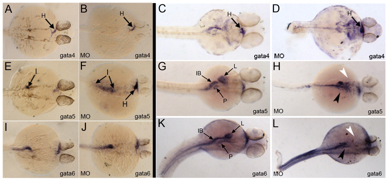
Fig. 5 Gata4 deficient fish are blocked at a relatively late stage of gut tube morphogenesis. Shown are representative examples of control embryos (A,C,E,G,I,K) and corresponding morphant (MO) embryos (B,D,F,H,J,L) following in situ hybridization using probes to detect Gata4 (A-D), Gata5 (E-H) or Gata6 (I-L). Embryos were fixed at 28 hpf (A,B,E,F,I,J) or 48 hpf (C,D,G,H,K,L). Note that, at 28 hpf, Gata5 expression is enhanced in the morphant embryos (arrows in F), whereas at 48 hpf Gata4 transcript levels are clearly increased (D), but Gata5 and Gata6 expression (H,L) is missing in the regions that normally show signal for pancreas (black arrowhead) and liver (white arrowhead). Views are dorsal, with anterior to the right. I, intestinal rod; H, heart; IB, intestinal bulb; L, liver; P, pancreas.