
- Title
-
Regulation of iro3 expression in the zebrafish spinal cord
- Authors
- Lewis, K.E., Bates, J., and Eisen, J.S.
- Source
- Full text @ Dev. Dyn.
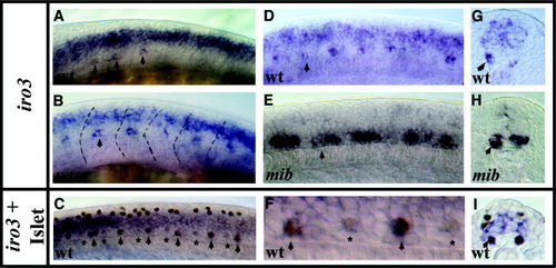
iro3 is expressed in CaP and VaP motoneurons. iro3 in situ hybridization (A,B,D,E,G,H) and Islet antibody staining + iro3 in situ hybridization (C,F,I). iro3 staining is blue and cytoplasmic; Islet antibody staining is brown and nuclear. Lateral views of the trunk at 6-8 somites (A), 10-12 somites (B), 18-20 somites (C-F), and cross-sections through the trunk at 18-20 somites (G-I). A-D,F,G,I: iro3 expression in wild-type (wt) embryos. E,H: iro3 expression in mib mutants. At all of these stages, in wild-type embryos, iro3 is expressed in a broad domain in intermediate spinal cord and in a subset of primary motoneurons (CaPs and VaPs). Arrows indicate iro3-expressing primary motoneurons (all of the iro3-expressing PMNs are indicated in A, C, and F, but only examples are indicated in B, D, E, and G-I); stars indicate primary motoneurons that do not yet express iro3 (MiPs and RoPs, brown only cells). The dotted lines in B demarcate somite boundaries. F is a close up of a two-somite length of the embryo shown in C. In mib mutants, the broad intermediate domain of iro3 expression is lost but additional cells in the primary motoneuron domain express iro3. Scale bar in A = 50 μm in A-E,G-I, 25 μm in F. EXPRESSION / LABELING:
|

iro3 is expressed by all motoneuron subtypes and by VeLD interneurons. Lateral trunk views of wild-type embryos at 24 hours postfertilization. Individual neurons were labeled with a mixture of fluorescein dextran and rhodamine dextran and then embryos with labeled neurons were processed for iro3 in situ hybridization (blue) and anti-fluorescein antibody staining (red). A-H: Shown are brightfield images (A-C,E,G) and fluorescent images (D,F,H). A: A labeled RoP that expresses iro3. B: A labeled SMN that expresses iro3. C,D: The same labeled CaP that expresses iro3. At this stage of development, CaP cell bodies have often moved dorsally, so that, like this example, they are now much closer to somite boundaries (Myers et al., [1986]). E,F: The same labeled MiP that expresses iro3. G,H: The same labeled VeLD that expresses iro3. In both A and C, the PMN is the more ventral of the two labeled cells and is indicated with white arrows; the more dorsal cells were inadvertently labeled by the pipette en route to the PMN. A,B: The labeled neuron was identified after the staining procedures by comparing its location with the previously recorded locations and identities of labeled cells in that embryo (see Experimental Procedures section). C: The white star indicates a small group of iro3-expressing ventral cells near the somite boundary (see text; this finding can be seen more clearly in Fig. 4A). Scale bar = 50 μm in A (applies to A-H). EXPRESSION / LABELING:
|
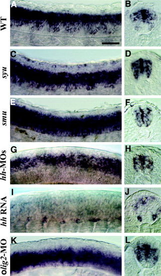
iro3 expression is regulated by Hedgehog (Hh) signals and Olig2. Lateral trunk views (A,C,E,G,I,K) and cross-sections through the trunk (B,D,F,H,J,L) showing iro3 in situ hybridization at 24 hours postfertilization (A-D) and 18-20 somites (E-L). A,B: Wild-type (WT) embryos. C,D: syu mutants. E,F: smu mutants. G,H: Wild-type embryos injected with hh-morpholino antisense oligonucleotides (MO), a severely affected embryo (G) and a slightly less severely affected embryo (H). I,J: Wild-type embryos injected with hh RNA. K,L: Wild-type embryos injected with olig2-MO. Dotted white lines in A demarcate somite boundaries. Scale bar = 50 μm in A (applies to A-L). EXPRESSION / LABELING:
|
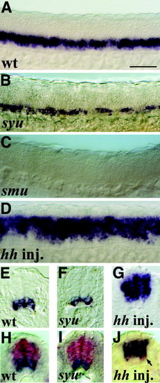
olig2 expression is regulated by Hh signals. Lateral trunk views (A-D) and cross-sections through the trunk (E-J) at 22-24 hours postfertilization showing olig2 single in situ hybridization (A-G) and olig2 (blue) and iro3 (red) double in situ hybridization (H-J). A,E,H: Wild-type (wt) embryos. B,F,I: syu mutants. C: smu mutant. D,G,J: Wild-type embryos injected with hh RNA. J: Most iro3 expression has been lost from the intermediate spinal cord, but iro3 is still expressed in motoneurons (example indicated with an arrow; see also Fig. 4I,J). Scale bar = 50 μm in A (applies to A-J). EXPRESSION / LABELING:
|