
FIGURE
Fig. 5
- ID
- ZDB-FIG-050204-10
- Publication
- Lewis et al., 2005 - Regulation of iro3 expression in the zebrafish spinal cord
- Other Figures
- All Figure Page
- Back to All Figure Page
Fig. 5
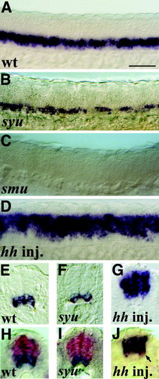
olig2 expression is regulated by Hh signals. Lateral trunk views (A-D) and cross-sections through the trunk (E-J) at 22-24 hours postfertilization showing olig2 single in situ hybridization (A-G) and olig2 (blue) and iro3 (red) double in situ hybridization (H-J). A,E,H: Wild-type (wt) embryos. B,F,I: syu mutants. C: smu mutant. D,G,J: Wild-type embryos injected with hh RNA. J: Most iro3 expression has been lost from the intermediate spinal cord, but iro3 is still expressed in motoneurons (example indicated with an arrow; see also Fig. 4I,J). Scale bar = 50 μm in A (applies to A-J). |
Expression Data
| Gene: | |
|---|---|
| Fish: | |
| Anatomical Term: | |
| Stage: | 26+ somites |
Expression Detail
Antibody Labeling
Phenotype Data
Phenotype Detail
Acknowledgments
This image is the copyrighted work of the attributed author or publisher, and
ZFIN has permission only to display this image to its users.
Additional permissions should be obtained from the applicable author or publisher of the image.
Full text @ Dev. Dyn.