
- Title
-
Neurogenin1 defines zebrafish cranial sensory Ganglia precursors
- Authors
- Andermann, P., Ungos, J., and Raible, D.W.
- Source
- Full text @ Dev. Biol.
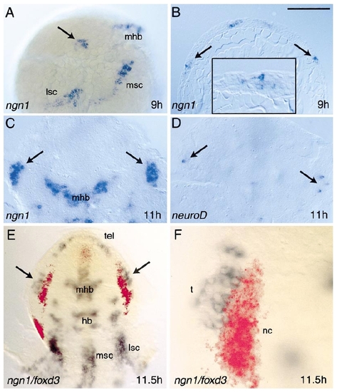
Development of trigeminal placodes. Trigeminal placodes first express ngn1 at 9 h (A, B). ngn1 expression appears nearly simultaneously within the trigeminal placode (arrows), and in patches of cells that will contribute to the mid/hindbrain (mhb), medial/ventral spinal cord (msc), and lateral/dorsal spinal cord (lsc). In (A), the embryo is shown in lateral view with anterior to the top and dorsal to the right. At this stage, the epiblast is several cell diameters thick and shows no distinct layering (B). ngn1-positive cells are found throughout the epiblast layer (B, inset). By 11 h, the trigeminal placodes have converged toward the midline (C) and begin to express neuroD (D). Embryos are flat-mounted dorsal view in (C,D) with anterior to the top. Relationship of the trigeminal placode with cranial neural crest (E, F). At 11.5 h, the trigeminal placode (arrows, t) expresses ngn1 (blue) and is located just lateral to the earliest differentiating neural crest cells (nc) marked with foxd3 (red). In (F), individual trigeminal cells can be distinguished adjacent to the neural crest. Embryo is shown in flat-mount dorsal view with anterior to the top. Bar = 200 μm (A, B, E); 50 μm (B, inset, F); 100 μm (C,D). |
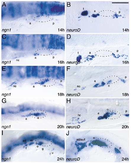
ngn1 and neuroD expression reveals the positions of the dorsolateral placodes. Panels show lateral views of zebrafish heads, just posterior to the eye, with anterior left and dorsal up. Neurogenic dorsolateral placodes form at the edges of the otic placode/vesicle, outlined with a dashed line. a, anterior lateral line placode area; ad, anterodorsal lateral line placode/ganglion; av, anteroventral lateral line placode/ganglion; f, facial epibranchial placode/ganglion; g, glossopharyngeal epibranchial placode/ganglion; m, middle lateral line placode/ganglion; nc, neural crest; o, octaval/statoacoustic ganglion precursors; p, posterior lateral line placode/ganglion; t, trigeminal placode/ganglion; v, vagal epibranchial placode/ganglion. Bar, 100 μm. |
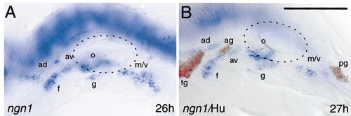
Spatial separation of anterior placodes. Anterior to the otic vesicle, ngn1-expressing anterodorsal, anteroventral, and facial placodes (blue) separate from an initially common area (see Fig. 2). The relationship of the placodes is apparent when embryos are stained with the anti-Hu antibody (red), which recognizes ganglion neurons. The otic vesicle is outlined with a dashed line. ad, anterodorsal lateral line placode; ag, anterodorsal lateral line ganglion; av, anteroventral lateral line placode; f, facial epibranchial placode/ganglion; g, glossopharyngeal epibranchial placode/ganglion; m, middle lateral line placode/ganglion; o, octaval/statoacoustic ganglion precursors; pg, posterior lateral line ganglion; tg, trigeminal ganglion; v, vagal epibranchial placode/ganglion. Bar, 100 μm. |
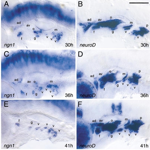
Development of epibranchial placodes. Each of the posterior pharyngeal pouches has an associated epibranchial placode, revealed by ngn1 expression. Cells continue to express neuroD after ngn1 is extinguished. ad, anterodorsal lateral line placode/ganglion; av, anteroventral lateral line placode/ganglion; f, facial epibranchial placode/ganglion; g, glossopharyngeal epibranchial placode/ganglion; m, middle lateral line placode/ganglion; o, octaval/statoacoustic ganglion precursors; v, vagal epibranchial placode/ganglion. Bar, 100 μm |
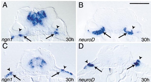
Relationship between ngn1 and neuroD expression in otic and vagal placodes. Transverse sections through the otic vesicle (A, B) and vagal placode (C, D) at 30 h are shown. ngn1 is expressed in the otic vesicle (arrowheads, A) and surface ectoderm (arrowheads, C), but down-regulated in cells soon after they delaminate (arrows). In contrast, fewer cells express neuroD in the otic vesicle (arrowhead, B) and surface ectoderm (arrowheads, D), but continue to express neuroD as cells coalesce into ganglia (arrows). Bar, 100 μm. |
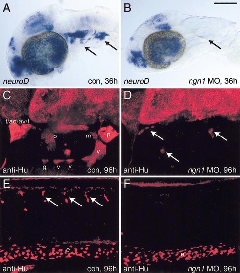
ngn1 morpholino oligonucleotides block differentiation of all cranial ganglia. Control embryos are shown in (A, C, E); ngn1 morpholino injected embryos in (B, D, F). (A, B) Lateral flat mounts of embryos expressing neuroD at 36 h. After ngn1 morpholino injection, neuroD expression is abolished in cranial ganglia (arrows) but not in other regions. (C, D) Lateral view of Hu expression in cranial ganglia at 96 h. In most cases, cranial ganglia are absent, but in some cases, a few neurons remain (arrows). (E, F) Lateral view of Hu expression in the zebrafish trunk at 96 h. The dorsal root ganglia (arrows) are eliminated by ngn1 morpholino injection, while enteric neurons are unaffected. ad, anterodorsal lateral line ganglion; av, anteroventral lateral line ganglion; e, enteric neurons; f, facial ganglion; g, glossopharyngeal ganglion; m, middle lateral line ganglion; o, octaval/statoacoustic ganglion; p, posterior lateral line ganglion; v, vagal ganglia. Bar, 100 μm (A, B) and (E, F); 30 μm (C, D). |
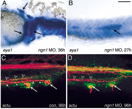
Lateral line development in the absence of ngn1 function. Migrating anterior lateral line (A) and posterior lateral line (B) primordia (arrows) form normally after injection of ngn1 morpholinos. Lateral line primordia are revealed by in situ hybridization for eya1. Injected embryos are indistinguishable from controls (not shown). Posterior lateral line primordia migrate to the tail and form normal neuromasts (C, D). The positions of neuromasts (arrows) are revealed by staining with anti-acetylated tubulin antibody (red) and sytox green (nuclear counterstain, green). Neuromasts are innervated by the posterior lateral line nerve (solid arrowheads) that in the tail is located just ventral and lateral to the ventral lateral fasiculus (outline arrowheads; Kuwada et al., 1990). Bar, 100 μm. |
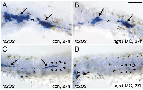
Glial cell differentiation after ngn1 morpholino injection. (A, B) Neural crest-derived glial cells initially differentiate despite the lack of cranial ganglia. In control embryos (A), glial cells found in association with cranial ganglia (arrows) on either side of the otic vesicle (ot) express foxd3. In injected embryos, glial cells are found in approximately the same positions, although sometimes foxd3 expression is lower. (C) Glial cells (arrows) are closely associated with the lateral line nerve and migrating primordium (outlined by arrowheads), but are missing after ngn1 morpholino injection. Bar, 100 μm. |
Reprinted from Developmental Biology, 251(1), Andermann, P., Ungos, J., and Raible, D.W., Neurogenin1 defines zebrafish cranial sensory Ganglia precursors, 45-58, Copyright (2002) with permission from Elsevier. Full text @ Dev. Biol.