
Fig. 1
- ID
- ZDB-FIG-080424-21
- Publication
- Andermann et al., 2002 - Neurogenin1 defines zebrafish cranial sensory Ganglia precursors
- Other Figures
- All Figure Page
- Back to All Figure Page
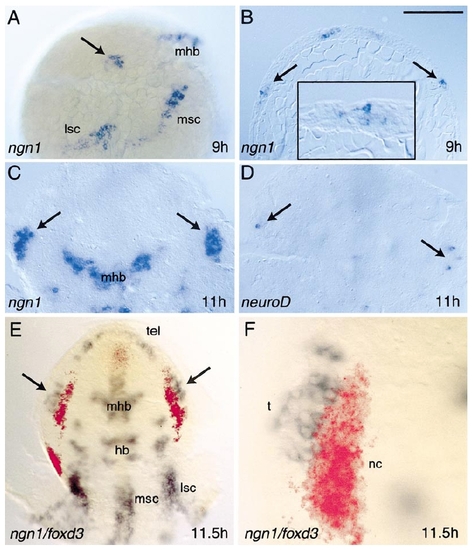
Development of trigeminal placodes. Trigeminal placodes first express ngn1 at 9 h (A, B). ngn1 expression appears nearly simultaneously within the trigeminal placode (arrows), and in patches of cells that will contribute to the mid/hindbrain (mhb), medial/ventral spinal cord (msc), and lateral/dorsal spinal cord (lsc). In (A), the embryo is shown in lateral view with anterior to the top and dorsal to the right. At this stage, the epiblast is several cell diameters thick and shows no distinct layering (B). ngn1-positive cells are found throughout the epiblast layer (B, inset). By 11 h, the trigeminal placodes have converged toward the midline (C) and begin to express neuroD (D). Embryos are flat-mounted dorsal view in (C,D) with anterior to the top. Relationship of the trigeminal placode with cranial neural crest (E, F). At 11.5 h, the trigeminal placode (arrows, t) expresses ngn1 (blue) and is located just lateral to the earliest differentiating neural crest cells (nc) marked with foxd3 (red). In (F), individual trigeminal cells can be distinguished adjacent to the neural crest. Embryo is shown in flat-mount dorsal view with anterior to the top. Bar = 200 μm (A, B, E); 50 μm (B, inset, F); 100 μm (C,D). |
Reprinted from Developmental Biology, 251(1), Andermann, P., Ungos, J., and Raible, D.W., Neurogenin1 defines zebrafish cranial sensory Ganglia precursors, 45-58, Copyright (2002) with permission from Elsevier. Full text @ Dev. Biol.