
Fig. 7
- ID
- ZDB-FIG-080424-27
- Publication
- Andermann et al., 2002 - Neurogenin1 defines zebrafish cranial sensory Ganglia precursors
- Other Figures
- All Figure Page
- Back to All Figure Page
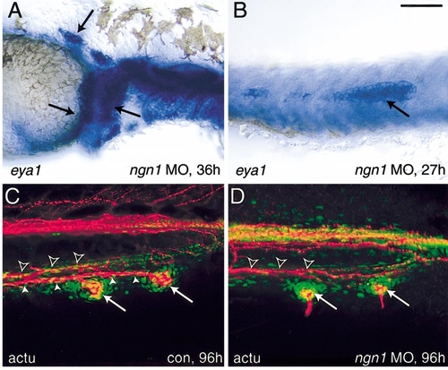
Lateral line development in the absence of ngn1 function. Migrating anterior lateral line (A) and posterior lateral line (B) primordia (arrows) form normally after injection of ngn1 morpholinos. Lateral line primordia are revealed by in situ hybridization for eya1. Injected embryos are indistinguishable from controls (not shown). Posterior lateral line primordia migrate to the tail and form normal neuromasts (C, D). The positions of neuromasts (arrows) are revealed by staining with anti-acetylated tubulin antibody (red) and sytox green (nuclear counterstain, green). Neuromasts are innervated by the posterior lateral line nerve (solid arrowheads) that in the tail is located just ventral and lateral to the ventral lateral fasiculus (outline arrowheads; Kuwada et al., 1990). Bar, 100 μm. |
Reprinted from Developmental Biology, 251(1), Andermann, P., Ungos, J., and Raible, D.W., Neurogenin1 defines zebrafish cranial sensory Ganglia precursors, 45-58, Copyright (2002) with permission from Elsevier. Full text @ Dev. Biol.