
|
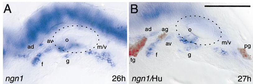
Fig. 3 Spatial separation of anterior placodes. Anterior to the otic vesicle, ngn1-expressing anterodorsal, anteroventral, and facial placodes (blue) separate from an initially common area (see Fig. 2). The relationship of the placodes is apparent when embryos are stained with the anti-Hu antibody (red), which recognizes ganglion neurons. The otic vesicle is outlined with a dashed line. ad, anterodorsal lateral line placode; ag, anterodorsal lateral line ganglion; av, anteroventral lateral line placode; f, facial epibranchial placode/ganglion; g, glossopharyngeal epibranchial placode/ganglion; m, middle lateral line placode/ganglion; o, octaval/statoacoustic ganglion precursors; pg, posterior lateral line ganglion; tg, trigeminal ganglion; v, vagal epibranchial placode/ganglion. Bar, 100 μm.
Reprinted from Developmental Biology, 251(1), Andermann, P., Ungos, J., and Raible, D.W., Neurogenin1 defines zebrafish cranial sensory Ganglia precursors, 45-58, Copyright (2002) with permission from Elsevier. Full text @ Dev. Biol.