
- Title
-
Midline morphogenesis of zebrafish foregut endoderm is dependent on Hoxb5b
- Authors
- Dalgin, G., Prince, V.E.
- Source
- Full text @ Dev. Biol.
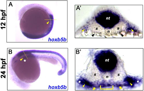
Hoxb5b is expressed in multiple tissues. (A–B) In situ hybridization for hoxb5b at 12 hpf and 24 hpf shows lateral plate mesoderm (LPM, yellow arrowhead) expression of hoxb5b transcripts (blue). (A′) Transverse section taken from the mid-trunk region of 12 hpf embryo in A (dashed yellow line) shows hoxb5b expression in neural tube (nt) and LPM regions (yellow arrowheads), low level sparse endoderm expression at or close to the limit of detection is indicated (black arrowheads). (B′) Transverse section taken from the mid-trunk region of 24 hpf embryo in B (dashed yellow line) shows hoxb5b expression in neural tube (nt), foregut endoderm (yellow bracket), adjacent mesenchyme (yellow arrows), and LPM (yellow arrowheads). The most lateral expression (white asterisks) is likely within the developing fin buds. Anterior to top (A) and left (B). |
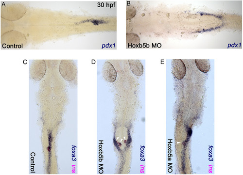
Hoxb5b morphants have bifurcated foregut endoderm. (A and C) Control embryos; (B and D) Hoxb5b morphants; (E) Hoxb5a morphant. (A and B) In situ hybridization for pdx1 and (C–E) double in situ hybridization for foxa3 (blue) and insulin (magenta) for control and morphant embryos at 30 hpf. Anterior to the left (A and B) and to the top (C–E), results are from 3 independent experiments with a minimum of 45 embryos for Hoxb5b morphants, and from 2 independent experiments with a minimum of 20 embryos for Hoxb5a morphants. |
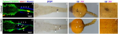
Hoxb5b morphants form endocrine pancreas cell types but fail to form liver cells. (A–C) Control embryos; (D–F) Hoxb5b morphants. (A and D) Confocal images of representative 30 hpf Tg (sox17:EGFP) embryos. Whole mount immunolabeling for GFP (green), Islet1 (red) and Myosin to label somites (blue) of control and Hoxb5b morphant embryos. (B and E) In situ hybridization for glucagon (gcga, blue); (C and F) double in situ hybridization for ceruloplasmin (cp, blue) and insulin (ins, magenta) for control and Hoxb5b morphant embryos at 30 hpf. (C′ and F′) high magnification views of foregut region. Anterior to the left, pancreatic cells (white arrowhead) and liver cells (black arrowhead). Results are from 2 independent experiments with a minimum of 25 embryos per group. Scale bar = 100 μm. |
Hoxb5b morphants form endocrine pancreas cell types but fail to form liver cells. (A–C) Control embryos; (D–F) Hoxb5b morphants. (A and D) Confocal images of representative 30 hpf Tg (sox17:EGFP) embryos. Whole mount immunolabeling for GFP (green), Islet1 (red) and Myosin to label somites (blue) of control and Hoxb5b morphant embryos. (B and E) In situ hybridization for glucagon (gcga, blue); (C and F) double in situ hybridization for ceruloplasmin (cp, blue) and insulin (ins, magenta) for control and Hoxb5b morphant embryos at 30 hpf. (C′ and F′) high magnification views of foregut region. Anterior to the left, pancreatic cells (white arrowhead) and liver cells (black arrowhead). Results are from 2 independent experiments with a minimum of 25 embryos per group. Scale bar = 100 μm. |
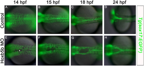
Midline defects in the foregut endoderm first become apparent at 14 hpf in Hoxb5b morphants. Fluorescent imaging of live Tg (sox17:EGFP) embryos (A–D) control, and (E–H) Hoxb5b morphant, at (A,E) 14 hpf; (B,F) 15 hpf; (C,G) 18 hpf; (D,H) 24 hpf. At 14 hpf endoderm cell-free patches are apparent at the midline in Hoxb5b-deficent specimens (asterisk). Anterior to the left, results are from 2 independent experiments and minimum of 8 embryos per group. |
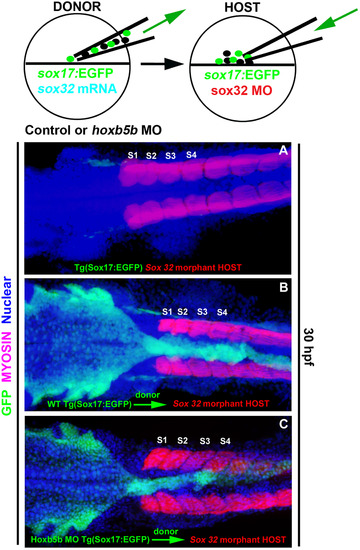
Hoxb5b function in the endoderm is not necessary for midline coalescence of the foregut endoderm. Cell transplantation approach: Tg (sox17:EGFP) embryos were used to follow the fate of endodermal cells in chimeric embryos. (A) Confocal image of a Sox32 morphant embryo; these lack endoderm and were used as hosts. (B) Tg (sox17:EGFP) donor embryos were injected with sox32 mRNA, or (C) together with Hoxb5b morpholino. Donor cells were transplanted into endoderm-deficient Sox32 morphant hosts. Whole mount immunolabeling for GFP (green), Myosin to label somites (magenta) and nuclear marker (blue). Both wild-type (WT) (B) and Hoxb5b morphant (C) donor cells reconstitute a normal gut in Sox32 morphant hosts (A). Anterior to the left, results are from 2 independent experiments with a minimum of 4 embryos per group. |
Reprinted from Developmental Biology, 471, Dalgin, G., Prince, V.E., Midline morphogenesis of zebrafish foregut endoderm is dependent on Hoxb5b, 1-9, Copyright (2020) with permission from Elsevier. Full text @ Dev. Biol.