
|
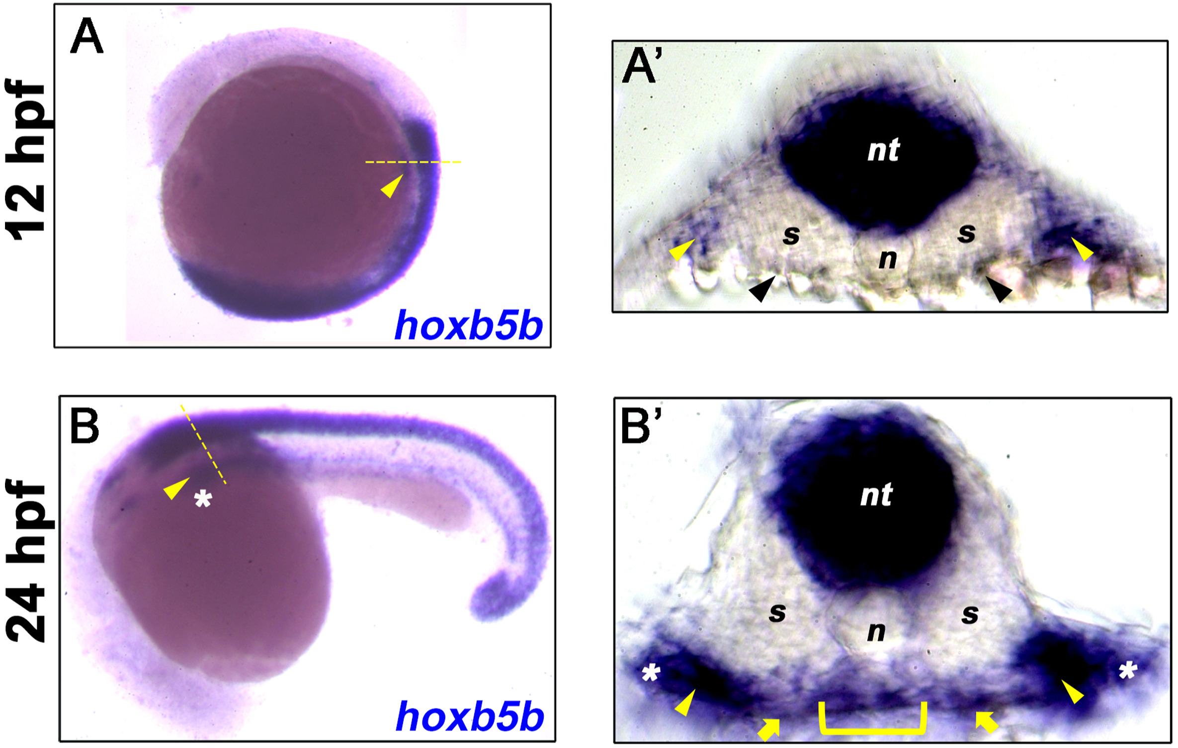
Fig. 1 Hoxb5b is expressed in multiple tissues. (A–B) In situ hybridization for hoxb5b at 12 hpf and 24 hpf shows lateral plate mesoderm (LPM, yellow arrowhead) expression of hoxb5b transcripts (blue). (A′) Transverse section taken from the mid-trunk region of 12 hpf embryo in A (dashed yellow line) shows hoxb5b expression in neural tube (nt) and LPM regions (yellow arrowheads), low level sparse endoderm expression at or close to the limit of detection is indicated (black arrowheads). (B′) Transverse section taken from the mid-trunk region of 24 hpf embryo in B (dashed yellow line) shows hoxb5b expression in neural tube (nt), foregut endoderm (yellow bracket), adjacent mesenchyme (yellow arrows), and LPM (yellow arrowheads). The most lateral expression (white asterisks) is likely within the developing fin buds. Anterior to top (A) and left (B).
Reprinted from Developmental Biology, 471, Dalgin, G., Prince, V.E., Midline morphogenesis of zebrafish foregut endoderm is dependent on Hoxb5b, 1-9, Copyright (2020) with permission from Elsevier. Full text @ Dev. Biol.