
Image
|
Figure Caption
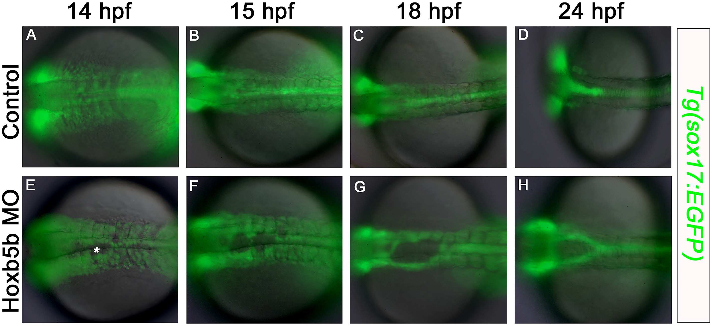
Fig. 5 Midline defects in the foregut endoderm first become apparent at 14 hpf in Hoxb5b morphants. Fluorescent imaging of live Tg (sox17:EGFP) embryos (A–D) control, and (E–H) Hoxb5b morphant, at (A,E) 14 hpf; (B,F) 15 hpf; (C,G) 18 hpf; (D,H) 24 hpf. At 14 hpf endoderm cell-free patches are apparent at the midline in Hoxb5b-deficent specimens (asterisk). Anterior to the left, results are from 2 independent experiments and minimum of 8 embryos per group.
Acknowledgments
This image is the copyrighted work of the attributed author or publisher, and
ZFIN has permission only to display this image to its users.
Additional permissions should be obtained from the applicable author or publisher of the image.
Reprinted from Developmental Biology, 471, Dalgin, G., Prince, V.E., Midline morphogenesis of zebrafish foregut endoderm is dependent on Hoxb5b, 1-9, Copyright (2020) with permission from Elsevier. Full text @ Dev. Biol.