- Title
-
Llgl1 regulates zebrafish cardiac development by mediating Yap stability in cardiomyocytes
- Authors
- Flinn, M.A., Otten, C., Brandt, Z.J., Bostrom, J.R., Kenarsary, A., Wan, T.C., Auchampach, J.A., Abdelilah-Seyfried, S., O'Meara, C.C., Link, B.A.
- Source
- Full text @ Development
|
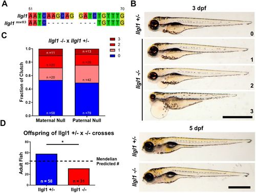
Assessment of llgl1 mutant gross morphology. (A) Diagram depicting the 10 bp deletion generated by TALENs in exon 1 of llgl1. (B) Embryos (3 dpf) from an llgl1+/−×llgl1−/− cross categorized into three grades of severity: class 0, wild-type phenotype; class 1 (mild), subtle signs of pericardial effusion at 3 dpf; class 2, pronounced pericardial effusion with a small subset displaying smaller eyes; and class 3 (severe), pronounced pericardial effusion and/or body edema, small eyes and hearts fail to undergo cardiac looping (Movie 5). At 5 dpf, llgl1−/− embryos are undistinguishable from llgl1+/− siblings. Scale bars: 1 mm. (C) Quantification of phenotype classes from llgl1+/−×llgl1−/− crosses comparing maternal contribution. Three clutches were used for each condition. n=109 for maternal llgl1−/− and n=160 for paternal llgl1−/−. χ2 test. (D) Adult genotyping from six pooled llgl1+/−×llgl1−/− crosses, greater than 4 months of age. The dashed line represents the predicted Mendelian number for each genotype. n=58 for llgl1+/− and n=31 for llgl1−/−. Two-tailed binomial test. Error bars indicate s.e.m. *P<0.05. PHENOTYPE:
|
|
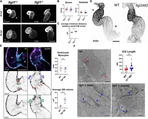
Loss of llgl1 disrupts normal heart development. (A) Representative confocal z-stack images of myl7:eGFP expression in hearts at 48 hpf and 99 hpf in llgl1+/− and llgl1−/− siblings. A, atrium; V, ventricle. (B) Quantification of atrial and ventricular cardiomyocyte nuclei in 48 hpf llgl1 morphants versus uninjected controls. n=9 for atrial nuclei analysis, n=6 for uninjected control and n=7 for llgl1 morpholino microinjection for ventricular nuclei analysis. (C) Quantification of average minimum distance between atrial cardiomyocyte nuclei in llgl1+/− and llgl1−/− siblings at 48 hpf. n=3 for llgl1+/− hearts and n=4 for llgl1−/− hearts. (D) Representative confocal z-stack images of actin-stained 48 hpf hearts. (E) Representative single-section confocal micrograph of alcam or actin-stained 48 hpf hearts, focusing on the AVC. Red arrows depict valve leaflets, blue arrows depict large rounded ventricular cardiomyocytes and green arrows depict abnormal actin staining in the myocardium. Quantification of ventricular cardiomyocyte number and volume. n=4 for untreated embryos and n=6 for llgl1 morpholino-treated embryos. (F) Representative electron micrographs of 4 dpf wild-type and llgl1−/− cardiomyocytes. Red asterisks denote normal sarcomeres of wild type, whereas blue asterisks denote thin and disorganized sarcomeres of llgl1−/− cardiomyocytes. Red arrows illustrate normal intercalated discs of wild type, whereas blue arrows indicate elongated dysmorphic intercalated discs of llgl1−/− cardiomyocytes. The hashtag indicates loss of adhesion between cardiomyocytes. Quantification of cardiomyocyte intercalated disc (ICD) length (top right). n=30 intercalated discs per group compiled from three animals per group, and ten micrographs per animal. Two-tailed unpaired Student's t-test. Error bars indicate s.e.m. ns, not significant, *P<0.05, **P<0.01. Scale bars: 25 μm (A); 50 μm (D,E); 500 nm (F). |

ZFIN is incorporating published figure images and captions as part of an ongoing project. Figures from some publications have not yet been curated, or are not available for display because of copyright restrictions. PHENOTYPE:
|
|
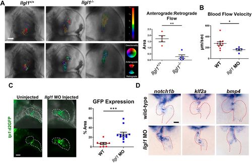
Valve morphology and hemodynamics are compromised in llgl1 mutants. (A) Representative images from videos acquired using light microscopy of 99 hpf hearts. A heart from an llgl1−/− embryo with slight pericardial effusion is displayed in the middle, whereas on the right, a heart with severe pericardial effusion is depicted. Above: blood flow was analyzed during diastasis using the particle image velocimetry in Fiji. Arrow vectors show directionality distance moved in pixels. Below: particle image velocimetry (PIV) data were analyzed using the PIV analyzer function in Fiji and qualified for anterograde or retrograde flow. Quantification of anterograde versus retrograde area within the heart chambers during diastasis is depicted in the graph (right). n=4 for llgl1+/+ embryos and n=5 for llgl1−/− embryos. (B) Blood flow velocity measurements analyzed from videos. n=10 for untreated embryos and n=5 for llgl1 morpholino-treated embryos. (C) Representative images from confocal z-stack micrographs of hearts of 2 dpf embryos carrying the tp1 transgenic Notch reporter. The area of cardiac GFP expression is depicted in the graph. n=9 for untreated embryos and n=12 for llgl1 morpholino-treated embryos. (D) Representative images of in situ hybridization of valve markers in 48 hpf fish. Ventral view focusing on the heart (red dashed line). Color model depicts the ventricle in red, atrium in blue, AVC in yellow and venous pole in purple. Two-tailed unpaired Student's t-test. Error bars indicate s.e.m. *P<0.05, **P<0.01, ***P<0.001. Scale bars: 100 µm. |
|
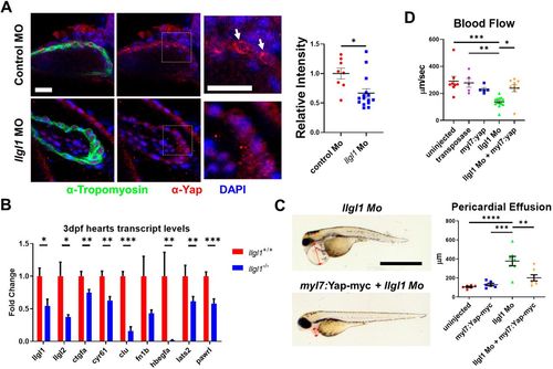
Llgl1 promotes appropriate Yap protein levels in cardiomyocytes, and exogenous expression of Yap in cardiomyocytes ameliorates deleterious cardiac physiology associated with loss of Llgl1. (A) Representative images of 2 dpf zebrafish ventricles from single plane confocal micrographs (left). Boxes indicate area of enlargement. Arrows depict Yap within the cardiomyocytes. Quantification of the intensity of anti-Yap signature in tropomyosin+ cells normalized to the control morpholino group (right). n=8 for untreated embryos and n=15 for llgl1 morpholino-treated embryos. (B) qRT-PCR transcript analysis for llgl1, llgl2 and genes regulated by Yap activity in llgl1+/+ or llgl1−/− 3 dpf zebrafish hearts. n=10 for each group. (C) Representative images of 3 dpf embryos microinjected with llgl1 morpholino with or without Tol2-mediated integration of myl7:yap-myc (left). Arrows depict the region measured for pericardial effusion. Quantification of pericardial effusion in 3 dpf embryos (right). n=6 for uninjected control group, n=6 for the myl7:yap-myc control group, n=7 for llgl1 morphants and n=7 for llgl1 morphants with myl7:yap-myc expression. (D) Quantification of blood flow velocity in 2 dpf embryos. n=8 for the uninjected control group, n=5 for the transposase only control group, n=5 for the myl7:yap-myc control group, n=13 for llgl1 morphants and n=9 for llgl1 morphants with myl7:yap-myc expression. Two-tailed unpaired Student's t test (A,B) and One-way ANOVA, Tukey's multiple comparisons test (C,D). Error bars indicate s.e.m. *P<0.05, **P<0.01, ***P<0.001,****P<0.0001. Scale bars: 50 µm (A); 1 mm (C). EXPRESSION / LABELING:
PHENOTYPE:
|

ZFIN is incorporating published figure images and captions as part of an ongoing project. Figures from some publications have not yet been curated, or are not available for display because of copyright restrictions. PHENOTYPE:
|

ZFIN is incorporating published figure images and captions as part of an ongoing project. Figures from some publications have not yet been curated, or are not available for display because of copyright restrictions. PHENOTYPE:
|

ZFIN is incorporating published figure images and captions as part of an ongoing project. Figures from some publications have not yet been curated, or are not available for display because of copyright restrictions. PHENOTYPE:
|

ZFIN is incorporating published figure images and captions as part of an ongoing project. Figures from some publications have not yet been curated, or are not available for display because of copyright restrictions. PHENOTYPE:
|

ZFIN is incorporating published figure images and captions as part of an ongoing project. Figures from some publications have not yet been curated, or are not available for display because of copyright restrictions. PHENOTYPE:
|

ZFIN is incorporating published figure images and captions as part of an ongoing project. Figures from some publications have not yet been curated, or are not available for display because of copyright restrictions. PHENOTYPE:
|

ZFIN is incorporating published figure images and captions as part of an ongoing project. Figures from some publications have not yet been curated, or are not available for display because of copyright restrictions. PHENOTYPE:
|

ZFIN is incorporating published figure images and captions as part of an ongoing project. Figures from some publications have not yet been curated, or are not available for display because of copyright restrictions. PHENOTYPE:
|

ZFIN is incorporating published figure images and captions as part of an ongoing project. Figures from some publications have not yet been curated, or are not available for display because of copyright restrictions. |

ZFIN is incorporating published figure images and captions as part of an ongoing project. Figures from some publications have not yet been curated, or are not available for display because of copyright restrictions. PHENOTYPE:
|


