
|
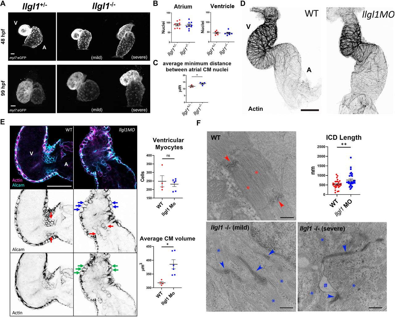
Fig. 3 Loss of llgl1 disrupts normal heart development. (A) Representative confocal z-stack images of myl7:eGFP expression in hearts at 48 hpf and 99 hpf in llgl1+/− and llgl1−/− siblings. A, atrium; V, ventricle. (B) Quantification of atrial and ventricular cardiomyocyte nuclei in 48 hpf llgl1 morphants versus uninjected controls. n=9 for atrial nuclei analysis, n=6 for uninjected control and n=7 for llgl1 morpholino microinjection for ventricular nuclei analysis. (C) Quantification of average minimum distance between atrial cardiomyocyte nuclei in llgl1+/− and llgl1−/− siblings at 48 hpf. n=3 for llgl1+/− hearts and n=4 for llgl1−/− hearts. (D) Representative confocal z-stack images of actin-stained 48 hpf hearts. (E) Representative single-section confocal micrograph of alcam or actin-stained 48 hpf hearts, focusing on the AVC. Red arrows depict valve leaflets, blue arrows depict large rounded ventricular cardiomyocytes and green arrows depict abnormal actin staining in the myocardium. Quantification of ventricular cardiomyocyte number and volume. n=4 for untreated embryos and n=6 for llgl1 morpholino-treated embryos. (F) Representative electron micrographs of 4 dpf wild-type and llgl1−/− cardiomyocytes. Red asterisks denote normal sarcomeres of wild type, whereas blue asterisks denote thin and disorganized sarcomeres of llgl1−/− cardiomyocytes. Red arrows illustrate normal intercalated discs of wild type, whereas blue arrows indicate elongated dysmorphic intercalated discs of llgl1−/− cardiomyocytes. The hashtag indicates loss of adhesion between cardiomyocytes. Quantification of cardiomyocyte intercalated disc (ICD) length (top right). n=30 intercalated discs per group compiled from three animals per group, and ten micrographs per animal. Two-tailed unpaired Student's t-test. Error bars indicate s.e.m. ns, not significant, *P<0.05, **P<0.01. Scale bars: 25 μm (A); 50 μm (D,E); 500 nm (F).