- Title
-
Dual-beam confocal light-sheet microscopy via flexible acousto-optic deflector
- Authors
- Gavryusev, V., Sancataldo, G., Ricci, P., Montalbano, A., Fornetto, C., Turrini, L., Laurino, A., Pesce, L., de Vito, G., Tiso, N., Vanzi, F., Silvestri, L., Pavone, F.S.
- Source
- Full text @ J. Biomed. Opt.
|
Image acquisition schemes: (a) in global shutter mode all pixels are exposed at once (orange color), while in (b) single- or (c) dual-rolling shutter modes only one or two sets of neighboring pixel rows are concurrently active, before sequentially enabling the next ones in the direction indicated by the arrows. The red line in (c) demarcates the sensor halves. |
|
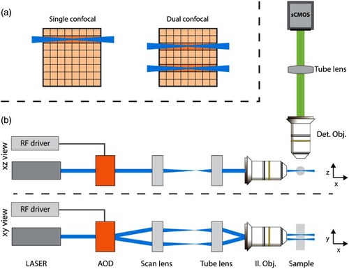
Schematic of: (a) sCMOS camera operating in single- or dual-rolling shutter mode with the illuminating beam (or beams) matching the position and synchronized with the scan rate of the virtual slit (or slits); (b) the excitation and imaging paths from side and top views. |
|
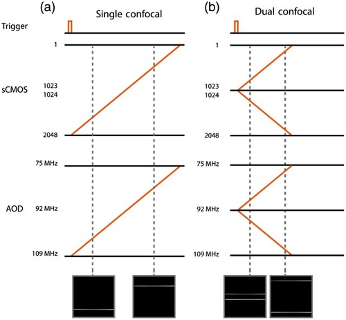
System timing configuration diagrams for single- (a) and dual-beam (b) confocal illumination. A common trigger starts the camera acquisition and tailored RF ramps on the signal generator that drives the AOD illumination sweep. The image insets are frames from Video |
|
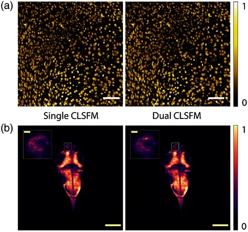
Representative single (right, top to bottom readout) and dual (left, diverging rolling shutter readout) beam CLSFM full-frame images of (a) cell nuclei in a mouse brain and (b) neuron nuclei in a zebrafish larva brain, respectively, color-coded in yellow and purple. The inset in (b) shows a four times magnified left habenula area within the diencephalon where neural activity can be observed. An extended dual CLSFM zebrafish brain time-lapse recording at 90 fps is shown in Video |
|
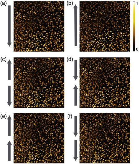
Representative (a) and (b) single and (c)–(f) dual beam CLSFM full-frame images of cell nuclei within the same mouse brain cortex area, acquired in the different rolling shutter readout direction modes of the sCMOS camera. No qualitative nor quantitative difference in the image quality is observable. |


