
FIGURE
Fig. 3
- ID
- ZDB-FIG-200212-16
- Publication
- Gavryusev et al., 2019 - Dual-beam confocal light-sheet microscopy via flexible acousto-optic deflector
- Other Figures
- All Figure Page
- Back to All Figure Page
Fig. 3
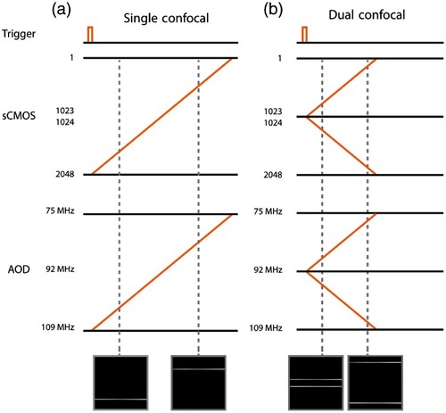
System timing configuration diagrams for single- (a) and dual-beam (b) confocal illumination. A common trigger starts the camera acquisition and tailored RF ramps on the signal generator that drives the AOD illumination sweep. The image insets are frames from Video |
Expression Data
Expression Detail
Antibody Labeling
Phenotype Data
Phenotype Detail
Acknowledgments
This image is the copyrighted work of the attributed author or publisher, and
ZFIN has permission only to display this image to its users.
Additional permissions should be obtained from the applicable author or publisher of the image.
Full text @ J. Biomed. Opt.