
Fig. 4
- ID
- ZDB-FIG-200212-24
- Publication
- Gavryusev et al., 2019 - Dual-beam confocal light-sheet microscopy via flexible acousto-optic deflector
- Other Figures
- All Figure Page
- Back to All Figure Page
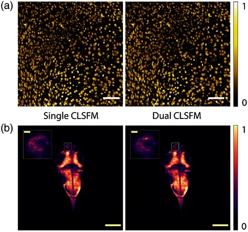
Representative single (right, top to bottom readout) and dual (left, diverging rolling shutter readout) beam CLSFM full-frame images of (a) cell nuclei in a mouse brain and (b) neuron nuclei in a zebrafish larva brain, respectively, color-coded in yellow and purple. The inset in (b) shows a four times magnified left habenula area within the diencephalon where neural activity can be observed. An extended dual CLSFM zebrafish brain time-lapse recording at 90 fps is shown in Video |