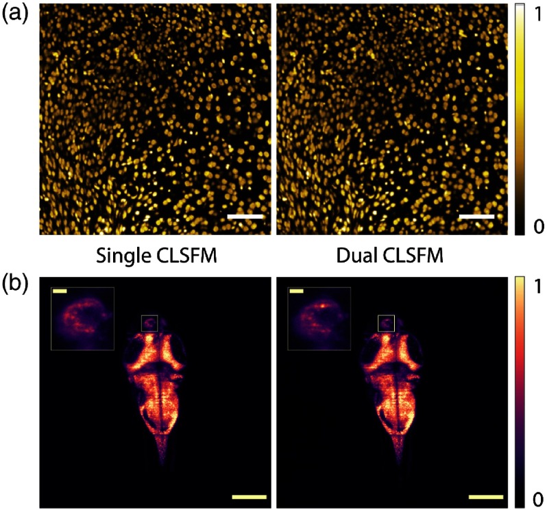
IMAGE

Fig. 4

ID
ZDB-IMAGE-200212-26
Source
Figures for Gavryusev et al., 2019
Image
Figure Caption

Fig. 4

Representative single (right, top to bottom readout) and dual (left, diverging rolling shutter readout) beam CLSFM full-frame images of (a) cell nuclei in a mouse brain and (b) neuron nuclei in a zebrafish larva brain, respectively, color-coded in yellow and purple. The inset in (b) shows a four times magnified left habenula area within the diencephalon where neural activity can be observed. An extended dual CLSFM zebrafish brain time-lapse recording at 90 fps is shown in Video 2 (MPEG, 2.8 MB [URL: https://doi.org/10.1117/1.JBO.24.10.106504.2], slowed down to 30 fps). Scale bar size: 200  μm; 20  μm in the inset.

Acknowledgments
This image is the copyrighted work of the attributed author or publisher, and ZFIN has permission only to display this image to its users. Additional permissions should be obtained from the applicable author or publisher of the image. Full text @ J. Biomed. Opt.