
Image
|
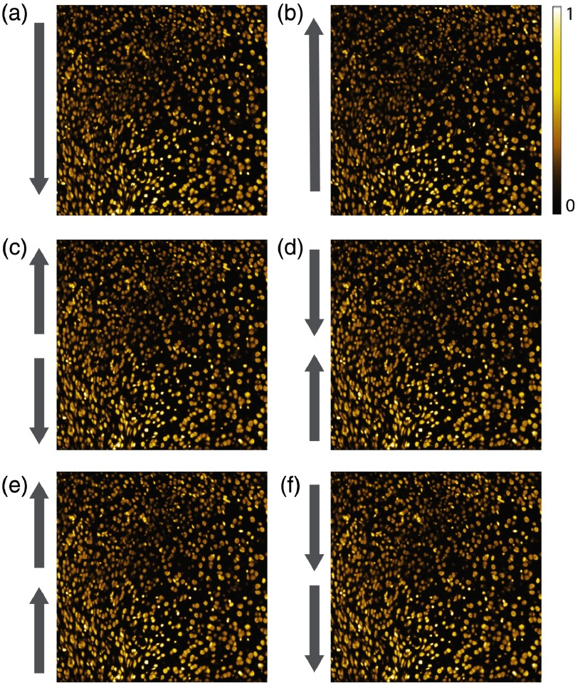
Figure Caption
Fig. 5
Representative (a) and (b) single and (c)–(f) dual beam CLSFM full-frame images of cell nuclei within the same mouse brain cortex area, acquired in the different rolling shutter readout direction modes of the sCMOS camera. No qualitative nor quantitative difference in the image quality is observable.
Acknowledgments
This image is the copyrighted work of the attributed author or publisher, and
ZFIN has permission only to display this image to its users.
Additional permissions should be obtained from the applicable author or publisher of the image.
Full text @ J. Biomed. Opt.