
- Title
-
Zebrafish Caudal Haematopoietic Embryonic Stromal Tissue (CHEST) Cells Support Haematopoiesis
- Authors
- Wolf, A., Aggio, J., Campbell, C., Wright, F., Marquez, G., Traver, D., Stachura, D.L.
- Source
- Full text @ Sci. Rep.
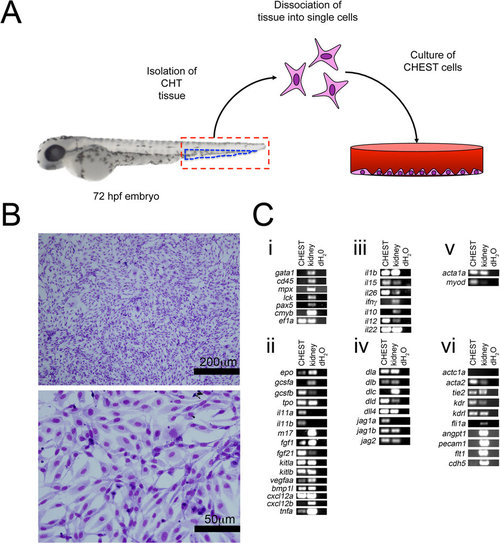
CHEST cells are a primary endothelial-like stromal cell line derived from the CHT of 72hpf zebrafish. (A) Isolation and culture strategy for CHEST cells. Red hatched lines indicate the tissue removed for deriving CHEST cells, and the blue hatched lines indicate the anatomical location of the CHT region. (B) Monolayers of CHEST cells stained with May-Grünwald Giemsa. Top image: 400x, scale bar is 200 μm; bottom image: 1000x, scale bar is 50 μm. (C) RT-PCR analysis of (i) hematopoietic, (ii) hematopoietic-supportive cytokines, (iii) inflammatory signals, (iv) Notch pathway mediators, (v) skeletal muscle, and (vi) cardiac and endothelial transcripts. Gel images have been cropped for clarity of presentation. |
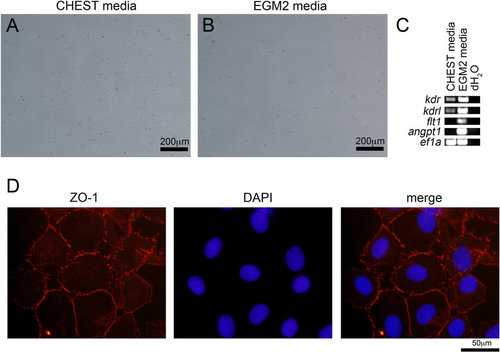
CHEST cells have endothelial-like properties. (A) Images of CHEST cells grown on tissue culture plates with CHEST growth media and (B) on Matrigel with EGM2 endothelial growth media. Images taken at 200x, scale bar is 200 μm. (C) RT-PCR performed on CHEST cells from (A) and (B) for endothelial-specific transcripts. (D) ZO-1 staining (red, left panel), DAPI staining (blue, middle panel), and merged image (right panel) of CHEST cells grown to confluence in CHEST media. Images taken at 1000x, scale bar is 50 μm. |
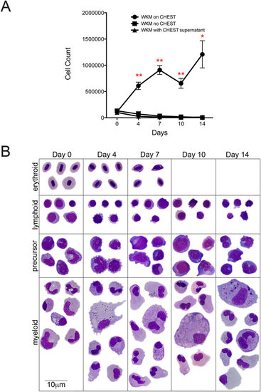
CHEST cells support hematopoietic expansion. (A) WKM from adult zebrafish was plated on CHEST cells and enumerated at various times (black circles; n = 8), plated in culture with no stromal under layer (black squares; n = 6), or plated with no CHEST cells in CHEST cell supernatant media (black triangles; n = 3). Error bars denote standard error of the mean. * denotes p < 0.005 and ** denotes p < 0.0005 when comparing cells grown on CHEST cells to either of the two other samples. (B) Composite May-Grünwald Giemsa images of hematopoietic cells isolated from the cultures (1000x, scale bar is 10 μm). |
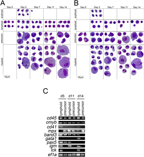
CHEST cells encourage differentiation of HSPCs in culture. (A) Composite May-Grünwald Giemsa images of hematopoietic cells isolated from the lymphoid fraction in Fig. 5 (1000x, scale bar is 10 μm). (B) Composite May-Grünwald Giemsa images of hematopoietic cells isolated from the precursor fraction in Fig. 5 (1000x, scale bar is 10 μm). (C) RT-PCR of leukocyte- (cd45), progenitor- (cmyb), erythromyeloid- (cd41, mpx, band3, and gata1), B cell- (pax5 and igm), and T cell- (lck) specific transcripts isolated from the cultures as in A and B. Gel images have been cropped for clarity of presentation. |