- Title
-
Functional visualization and disruption of targeted genes using CRISPR/Cas9-mediated eGFP reporter integration in zebrafish
- Authors
- Ota, S., Taimatsu, K., Yanagi, K., Namiki, T., Ohga, R., Higashijima, S.I., Kawahara, A.
- Source
- Full text @ Sci. Rep.
|
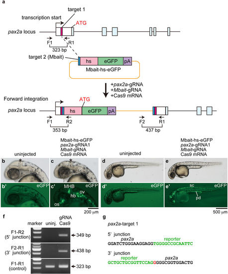
Targeted genomic integration of the Mbait-hs-eGFP reporter into the pax2a locus using the CRISPR/Cas9. (a) A schematic representation of the pax2a locus containing the 5′ untranslated region (white box) and the pax2a-gRNA target sites (pink box), and the reporter construct consisting of the Mbait-gRNA target site (blue box), the hsp70 promoter (hs, light red box), the eGFP gene (green box) and the polyA signal (pA, purple box). The Mbait-hs-eGFP reporter was co-injected with pax2a-gRNA1, Mbait-gRNA and Cas9 mRNA into 1-cell stage zebrafish embryos. Concurrent cleavage of the targeted genomic locus and the Mbait-hs-eGFP reporter resulted in the integration of the reporter by non-homologous end joining. The scheme shows forward integration of the reporter. (b,b’,d,d’) An uninjected control embryo around 32 hpf shows no eGFP expression. (c,c’,e,e’) The expression of eGFP around 32 hpf was detected in the midbrain-hindbrain boundary (MHB)(asterisk), the optic stalk (os), the otic vesicles (ov), the hindbrain neurons (hb), the spinal cord (sc) neurons and the pronephric duct (pd) in the injected embryo at 1 dpf. (f) The integration of the reporter into the pax2a gene was determined by genomic PCR using the pax2a-specific and reporter-specific primers. The targeted positions of the primers are shown in (a). (g) The sequences of the 5′ junction and the 3′ junction at the integration site of the eGFP-positive embryo (c,c’). A 4-bp deletion and a 1-bp insertion were observed at the 5′ junction and the 3′ junction, respectively. The red letter represents the inserted nucleotide. (b–e,b’–e’) Lateral view with anterior to the left and dorsal to the top. Black letters and green letters represent pax2a sequences and the reporter sequences, respectively. |
|
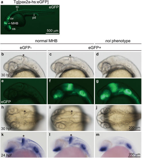
eGFP expression and noi phenotype in Tg[pax2a-hs:eGFP] embryos. (a) A heterozygous Tg[pax2a-hs:eGFP] embryo at 30 hpf. The expression of eGFP was detected in the MHB, the optic stalk (os), the otic vesicles (ov), the spinal cord (sc) neurons and the pronephric duct (pd). (b–j) The 30 hpf F2 embryos derived from mating of heterozygous Tg[pax2a-hs:eGFP] F1 fish. (b,e,h) A wild-type embryo. The isthmus is formed at the MHB (asterisk). (c,f,i) A heterozygous Tg[pax2a-hs:eGFP] embryo. The isthmus is formed normally. (d,g,j) A homozygous Tg[pax2a-hs:eGFP] embryo. eGFP expression around the MHB was anteriorly expanded. The isthmus was not formed in the homozygous Tg[pax2a-hs:eGFP] embryo. Genotyping was performed using genomic PCR. (k–m) Whole-mount in situ hybridization using anti-sense pax2a RNA probe at 24 hpf. The pax2a expression in the MHB was detected in the wild-type (k) and the heterozygous (l) embryos, but not in the homozygous embryos (m). (a–g,k–m) Lateral view with anterior to the left and dorsal to the top. (h–j) Dorsal view with anterior to the left. |
|
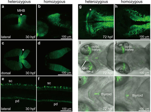
eGFP expression visualized by the endogenous pax2a enhancer activity. (a,c) The MHB (asterisk) of a heterozygous Tg[pax2a-hs:eGFP] embryo at 30 hpf. (b,d) The midbrain and hindbrain of a homozygous Tg[pax2a-hs:eGFP] embryo at 30 hpf. (e,f) The spinal cord neurons (sc) and pronephric duct (pd) of heterozygous (e) and homozygous (f) Tg[pax2a-hs:eGFP] embryos at 30 hpf. (g) The MHB of the heterozygous Tg[pax2a-hs:eGFP] embryo at 72 hpf. (h) The midbrain and hindbrain of the homozygous Tg[pax2a-hs:eGFP] embryo at 72 hpf. (i,j) The optic nerves of heterozygous (i) and homozygous (j) Tg[pax2a-hs:eGFP] embryos at 72 hpf. (k,l) The thyroids of heterozygous (k) and homozygous (l) Tg[pax2a-hs:eGFP] embryos at 72 hpf. All data were obtained by confocal microscopy. (a,b,e,f) Genotyping was performed using genomic PCR. Lateral view with anterior to the left and dorsal to the top. (c,d,g,h) Dorsal view with anterior to the left. (i–l) Ventral view with anterior to the left. EXPRESSION / LABELING:
PHENOTYPE:
|
|
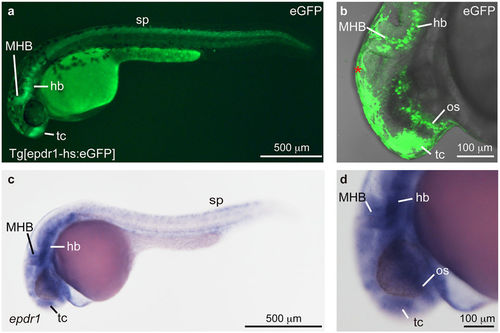
Establishment of Tg[epdr1-hs:eGFP]. (a,b) A heterozygous Tg[epdr1-hs:eGFP] embryo. eGFP expression was detected in the telencephalon (tc), the midbrain-hindbrain boundary (MHB), the hindbrain (hb) and spinal neurons (sp) at 30 hpf. The epdr1 expression in the optic stalk (os) was detected by confocal microscope (b). Autofluorescence of epidermis was detected (*). (c,d) Expression of epdr1 in the tc, the os, the MHB, the hb and the sp at 24 hpf. The expression of epdr1 was examined by whole-mount in situ hybridization using an anti-sense epdr1 RNA probe. |
|
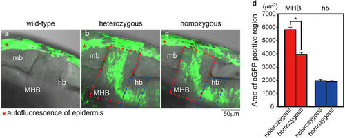
Functional analysis of Tg[epdr1-hs:eGFP]. (a) Autofluorescence of epidermis in wild-type embryo (red asterisk). (b,c) The distribution of eGFP-positive cells in the midbrain (mb) and the hindbrain (hb) of heterozygous (b) and homozygous (c) Tg[epdr1-hs:eGFP] embryos at 30 hpf (lateral view, anterior left) (confocal images). Autofluorescence in epidermis is indicated by red asterisk. (d) The area of eGFP-positive cells of heterozygous and homozygous Tg[epdr1-hs:eGFP] embryos in the MHB (red dashed line) and anterior part of hindbrain (blue dashed line) in the visual field (N = 14 each). Error bars indicate standard error of the mean (SEM). Statistical significance was determined using Student’s t-test. *P < 0.05. |
|
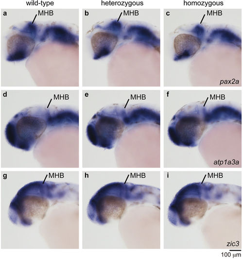
Expression of neural genes in the midbrain and the hindbrain of Tg[epdr1-hs:eGFP] embryos. The expression of neural marker genes was examined by whole-mount in situ hybridization using the indicated anti-sense RNA probes at 30 hpf. The expression of pax2a in the MHB and the optic stalk of the homozygous Tg[epdr1-hs:eGFP] embryo was slightly decreased compared with that of the wild-type or the heterozygous Tg[epdr1-hs:eGFP] embryos. The expression of atp1a3a and zic3 was not affected. |
|
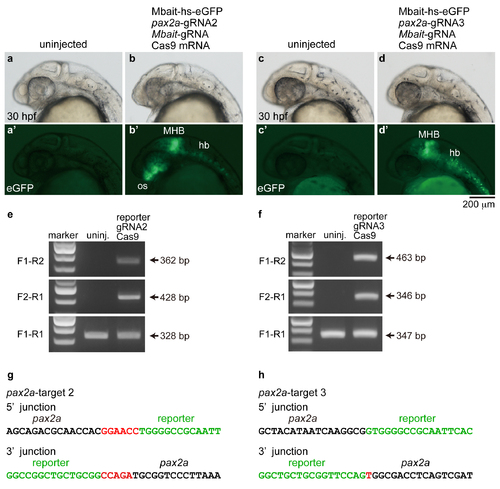
Targeted genomic integration of the Mbait-hs-eGFP into the pax2a locus using the CRISPR/Cas9. (a, a', c, c') Uninjected embryos. (b, b') An embryo injected with the reporter, pax2a-gRNA2, Mbait-gRNA and Cas9 mRNA. The expression of eGFP was detected in the MHB, the optic stalk (os) and the hindbrain neurons (hb). (d, d') An embryo injected with the reporter, pax2a-gRNA3, Mbait-gRNA and Cas9 mRNA. The expression of eGFP was detected in the MHB and the hindbrain neurons. (e, f) The integrations of the reporter in the pax2a gene (e; pax2a-target 2, f; pax2a-target 3) were determined by genomic PCR using the pax2a-specific and reporter-specific primers. Targeted positions of the primers are shown in Figure 1. (g, h) Genomic sequence of the 5' junction and the 3' junction at the integration site in the eGFP-positive embryo. Inserted nucleotides are indicated in red letters. Black letters and green letters represent pax2a sequences and the reporter sequences, respectively. |
|
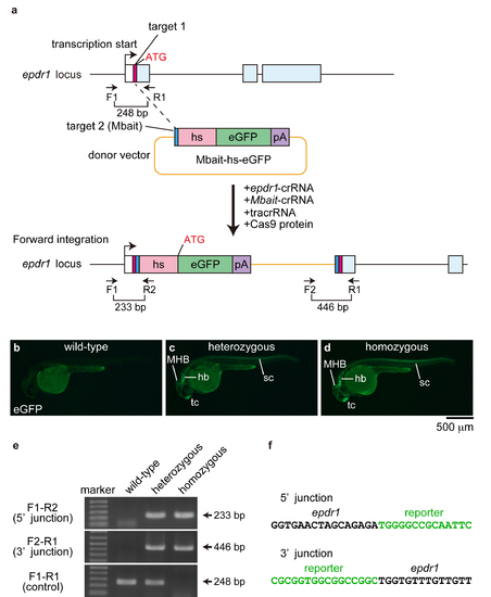
The eGFP expression in Tg[epdr1-hs:eGFP] embryos. (a) A schematic representation of the epdr1 locus containing the 5' untranslated region (white box) and the epdr1-crRNA target site (pink box), and the reporter construct consisting of the Mbait-crRNA target site (blue box), the hsp70 promoter (hs, light red box), the eGFP gene (green box) and the polyA signal (pA; purple box). (b-d) eGFP expression in wild-type, heterozygous and homozygous Tg[epdr1-hs:eGFP] embryos. (e) The integration of the reporter into the epdr1 locus was determined by genomic PCR using the epdr1-specific and the reporter-specific primers. (f) Sequence of the 5' junction and the 3' junction at the integration site of Tg[epdr1-hs:eGFP]. Black letters and green letters represent epdr1 sequences and reporter sequences, respectively. |

