
|
Fig. S2
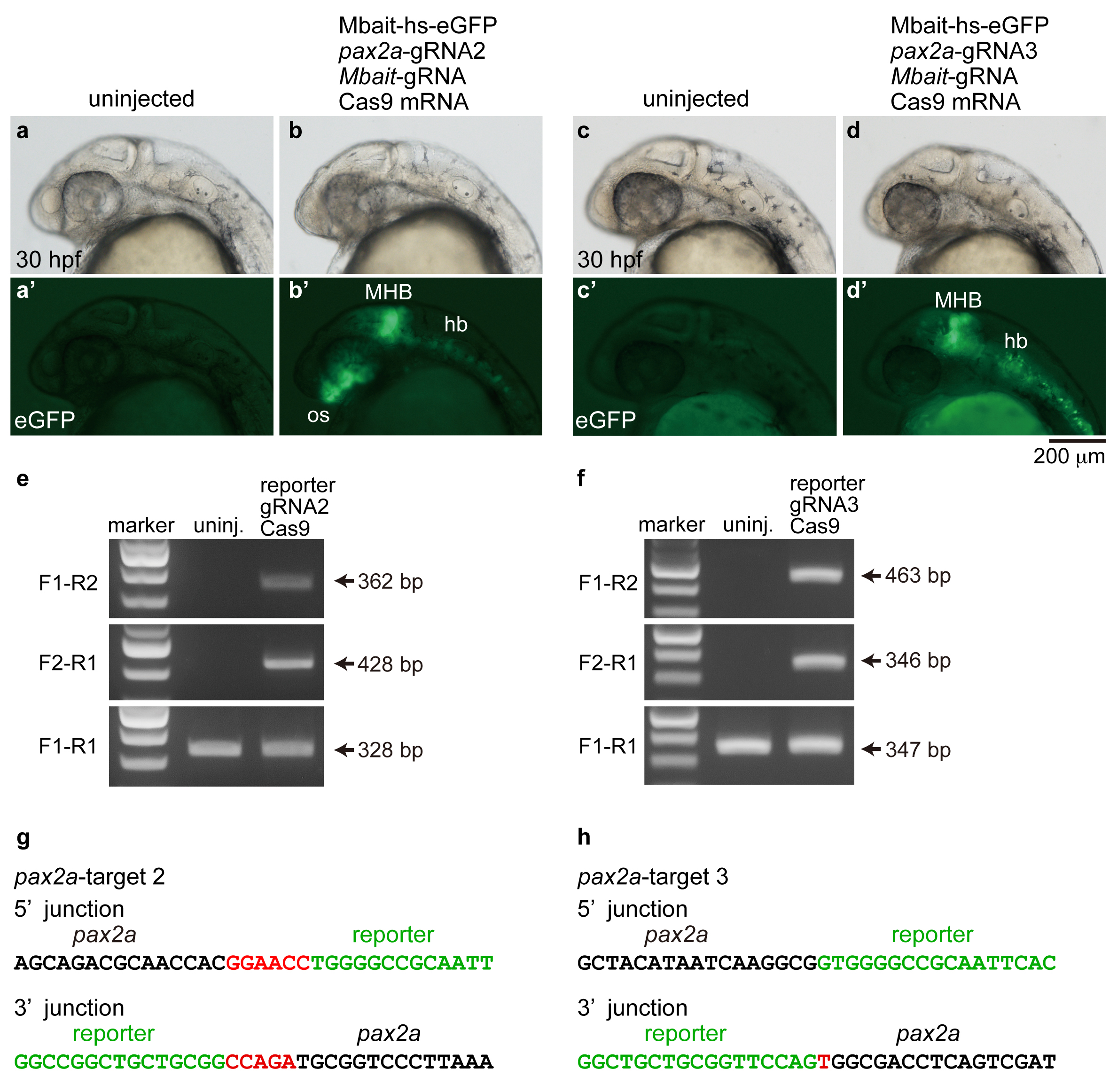
Targeted genomic integration of the Mbait-hs-eGFP into the pax2a locus using the CRISPR/Cas9.
(a, a', c, c') Uninjected embryos. (b, b') An embryo injected with the reporter, pax2a-gRNA2, Mbait-gRNA and Cas9 mRNA. The expression of eGFP was detected in the MHB, the optic stalk (os) and the hindbrain neurons (hb). (d, d') An embryo injected with the reporter, pax2a-gRNA3, Mbait-gRNA and Cas9 mRNA. The expression of eGFP was detected in the MHB and the hindbrain neurons. (e, f) The integrations of the reporter in the pax2a gene (e; pax2a-target 2, f; pax2a-target 3) were determined by genomic PCR using the pax2a-specific and reporter-specific primers. Targeted positions of the primers are shown in Figure 1. (g, h) Genomic sequence of the 5' junction and the 3' junction at the integration site in the eGFP-positive embryo. Inserted nucleotides are indicated in red letters. Black letters and green letters represent pax2a sequences and the reporter sequences, respectively.