
- Title
-
Kinesin family member 6 (kif6) is necessary for spine development in zebrafish
- Authors
- Buchan, J.G., Gray, R.S., Gansner, J.M., Alvarado, D.M., Burgert, L., Gitlin, J.D., Gurnett, C.A., Goldsmith, M.I.
- Source
- Full text @ Dev. Dyn.
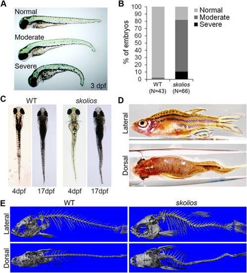
Skolios mutants develop recessively inherited curvature of the body axis. A: Ventral curvature phenotypes observed in skolios mutant embryos at 3 dpf. B: Relative frequencies of ventral curvature phenotypes in WT and skolios embryos (3 dpf). C: Dorsal view of embryonic (4 dpf) and juvenile (17 dpf) zebrafish showing development of sagittal body curvature in skolios mutants. D: Severe body curvature in both the medial–lateral and dorsal–ventral planes of an adult skolios mutant. E: MicroCT imaging at approximately 50 dpf shows marked curvature of the abdominal (rib vertebrae) and caudal (vertebrae posterior to ribs) spine in skolios mutants. PHENOTYPE:
|

Spinal curvature in skolios mutants occurs independent of major vertebral abnormalities and is progressive through adult stages in males. A: Method of measuring spinal curve in zebrafish using skeletal histomorphology. Lines were drawn through the middle of the two vertebrae adjacent to the apex of the curve. The measured angle from the intersection was subtracted from 180° to calculate the spinal curve. B: Spinal curves measured for female skolios mutants at 2 (N = 11), 5 (N = 9), 7 (N = 5) and 18 (N = 5) mpf and male skolios mutants at 2 (N = 10), 5 (N = 8), 7 (N = 5) and 18 (N = 5) mpf. The average spinal curve is shown for each time point. At 2 mpf, males and females are similarly affected; however, by later stages, males continue to progress in severity. *P < 0.05. C: Skeletal histomorphology of a 2 mpf female WT and a skolios mutant with a 35° spinal curve near the transition from abdominal to caudal vertebrae that developed in the absence of vertebral fusions or other major vertebral abnormalities. Box identifies region shown in A. D: Zebrafish between 2 and 7 mpf were stained with Alizarin red to visualize individual vertebrae. Staining shows vertebral fusions (arrows) identified in WT and skolios mutants. E: The overall frequency of vertebral fusions in skolios mutants is similar to WT. PHENOTYPE:
|
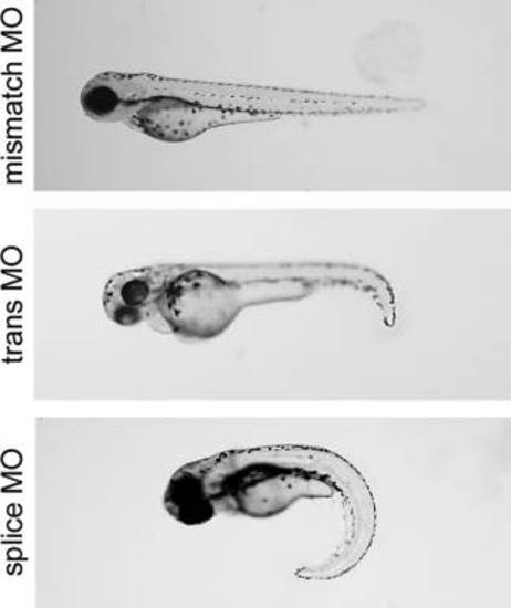
Transient knockdown of kif6 using morpholinos. WT zebrafish were injected with morpholinos targeting either the translation start site or a splice site, at the 1–4 cell stage. A morpholino targeting the kif6 translation start site but containing a 5 base mismatch (mismatch MO) served as a negative control. Both translation start site and splice site morphants recapitulated the early embryonic curvature of skolios. PHENOTYPE:
|
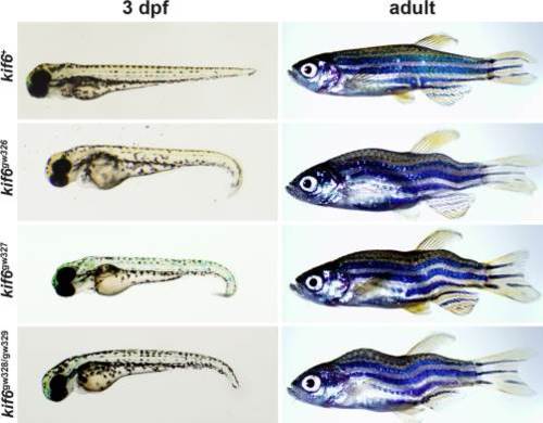
TALEN-induced mutations in kif6 recapitulate the skolios phenotype. Zebrafish with TALEN-induced mutations in kif6 (gw327, gw328, gw329) develop both embryonic and adult body axis curvature, similar to kif6gw326 mutants. PHENOTYPE:
|
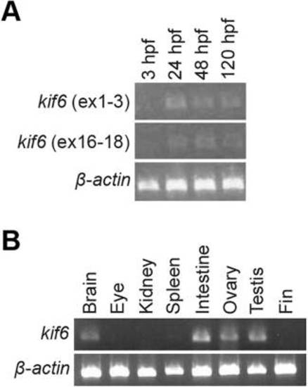
RT-PCR expression of kif6 in WT embryos and WT adult tissues. A: RT-PCR in WT zebrafish embryos shows low levels of kif6 between 24 and 120 hpf. B: RT-PCR of zebrafish tissues shows kif6 expression primarily in brain, intestine and reproductive organs. |
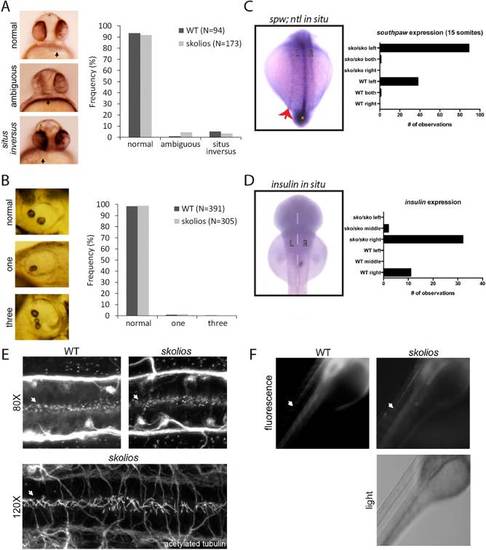
Cilia structure and function are normal in kif6gw326 mutants. A: Defects in left–right asymmetry were evaluated by visualizing the position of the heart in 30 hpf embryos for WT (N = 94) and kif6gw326 mutants (N = 173). Normally, zebrafish hearts are located to the left of the midline (right side when viewed ventrally) at 30 hpf. In zebrafish with ambiguous heart positioning, hearts appear at the midline and to the right side (left side when viewed ventrally) in zebrafish with situs inversus. Ambiguous and inversed orientations of the heart were observed at similar frequencies in WT and kif6gw326 embryos. B: Otoliths were observed laterally in 48–72 hpf zebrafish embryos. Normal otolith numbers (two) and abnormal otolith numbers (one or three) were quantified WT (N = 371) and kif6gw326 mutants (N = 305). C: In situ hybridization for southpaw (spw) to highlight LR patterning and no tail (ntl) to highlight the midline shows no defect in left–right patterning in kif6 sko/sko mutants (n = 39 WT and 90 kif6 sko/sko mutants). D: In situ hybridization for insulin (ins) to highlight LR patterning in the pancreas shows no defect in the normally rightward place of the pancreas in kif6 sko/sko mutants (n = 11 WT and 34 kif6 sko/sko mutants). E: Whole-mount confocal immunostaining of 28 hpf WT and kif6gw326 mutant embryos shows normal appearance of cilial in the central canal (arrows). F: Live 48 hpf WT and kif6gw326 embryos were injected with 5% tetramethylrhodamine conjugated to 70,000 molecular weight dextran. Sixty minutes after injection, the dye has migrated similar distances in skolios mutants compared with WT embryos. EXPRESSION / LABELING:
PHENOTYPE:
|