
|
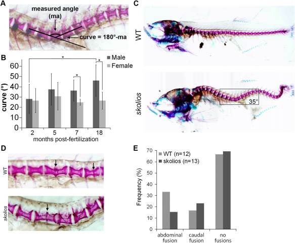
Fig. 2 Spinal curvature in skolios mutants occurs independent of major vertebral abnormalities and is progressive through adult stages in males. A: Method of measuring spinal curve in zebrafish using skeletal histomorphology. Lines were drawn through the middle of the two vertebrae adjacent to the apex of the curve. The measured angle from the intersection was subtracted from 180° to calculate the spinal curve. B: Spinal curves measured for female skolios mutants at 2 (N = 11), 5 (N = 9), 7 (N = 5) and 18 (N = 5) mpf and male skolios mutants at 2 (N = 10), 5 (N = 8), 7 (N = 5) and 18 (N = 5) mpf. The average spinal curve is shown for each time point. At 2 mpf, males and females are similarly affected; however, by later stages, males continue to progress in severity. *P < 0.05. C: Skeletal histomorphology of a 2 mpf female WT and a skolios mutant with a 35° spinal curve near the transition from abdominal to caudal vertebrae that developed in the absence of vertebral fusions or other major vertebral abnormalities. Box identifies region shown in A. D: Zebrafish between 2 and 7 mpf were stained with Alizarin red to visualize individual vertebrae. Staining shows vertebral fusions (arrows) identified in WT and skolios mutants. E: The overall frequency of vertebral fusions in skolios mutants is similar to WT.