
- Title
-
edn1 and hand2 Interact in Early Regulation of Pharyngeal Arch Outgrowth during Zebrafish Development
- Authors
- Sasaki, M.M., Nichols, J.T., and Kimmel, C.B.
- Source
- Full text @ PLoS One
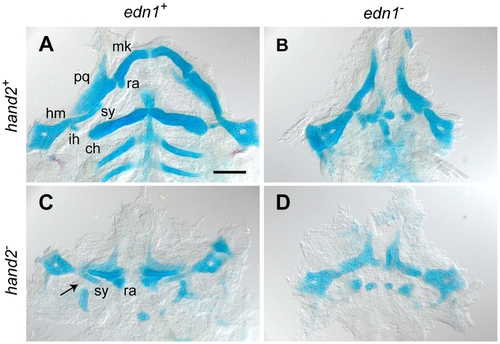
edn1 and hand2 are required for ventral jaw cartilage development. 4 dpf zebrafish skeletons were cartilage and bone stained with Alcian Blue and Alizarin Red, dissected and flat-mounted. (A) WT, with cartilages of the first two arches indicated: In the first arch the dorsal palatoquadrate (pq) and ventral Meckel’s cartilage (mk) articulate, Meckel’s cartilage includes a distinctive retroarticular process (ra) adjacent to the joint. In the second arch the more dorsal hyosymplectic cartilage is subdivided into the hyomandibular (hm) and symplectic (sy) regions, and the prominent ventral cartilage is the ceratohyal (ch). The interhyal cartilage (ih) forms a small hinge within the joint region. (B) In the edn1 larva, ventral cartilages and elements of the joint regions are missing or prominently disrupted. Unidentifiable elements are scattered near the ventral midline. (C) The hand2 larva exhibits ventral reductions similar to edn1, but the arch 2 symplectic cartilage and joint (arrow) are present and the arch 1 retroarticular process is expanded rather than missing. (D) The edn1;hand2 larva exhibits defects similar to edn1; in particular, the symplectic cartilage and retroarticular process cannot be identified. Anterior is upward and right is towards the left. Scale bar: 100 μm. |
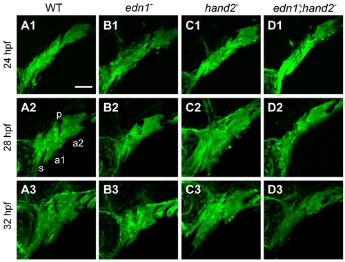
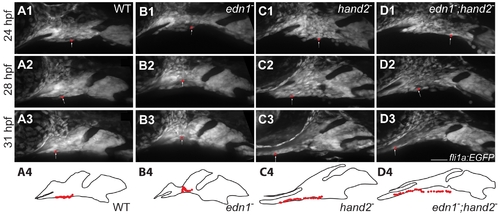
edn1 and hand2 are required for proper growth and morphogenesis of the early pharyngeal arches. Projections of lateral confocal images of embryos expressing fli1a:EGFP taken at 24, 28, and 32 hpf. This figure provides an overview of changes in the first two arches (a1, a2, separated by the first pharyngeal pouch, p) that are quantified and more fully documented in subsequent figures. Dorsal is to the top in each panel, and anterior is to the left. No differences in arch morphology are observed among the four genotypes at 24 hpf (upper row of panels). Subsequently the arches prominently lengthen along the DV axis; this DV extension is evidently reduced in the edn1 mutant (B2,B3) and edn1;hand2 double mutant (D2,D3) compared to WT and the hand2 mutant. The arches (particularly the first), also shorten along the AP axis but there appears to be no differences in AP shortening among the genotypes. The hand2 mutant also shows marked expansion of mesenchyme ventral to the stomodeum (s) in the anterior first arch (C2,C3). Scale bar: 50 μm. |
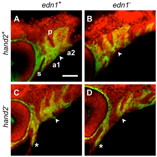
Counterstaining the fli1a:EGFP-expressing pharyngeal arches (a1, a2) with the nucleic acid stain SYTO 59 (red) clarifies the ectomesenchymal phenotypes (double-labeled). The DV ventral extension differences (arrowheads) are apparent ventral to the red-stained first pharyngeal pouch (p). Compared to the WT (A) this ventral region of mesenchyme is reduced in the edn1 mutant (B) and double mutant (D), and expanded in the hand2 mutant (C). Anterior extension of mesenchyme ventral to the stomodeum (s) is prominent in the hand2 mutant and double mutant (*, C, D). |
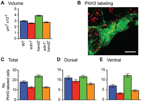
edn1 positively and hand2 negatively regulate the volume of pharyngeal arch fli1a:EGFP expressing mesenchyme (A) and ventral domain proliferation of neural crest derived cells (B–E). Data are for 28 hpf, a minimum of 23 individuals of each genotype were sampled. (B) shows an example 2-color image of phospho-histone H3 labeling for mitosing cells (red) and fli1a:EGFP expression (green). (C–E) show mean counts of double-labeled cells ±SEM. Tukey-Kramer comparison of the mean volumes (in A) reveals that the edn1 mutant volume is significantly lower than WT, and that the hand2 mutant volume is significantly higher (P<0.05). Statistically the double mutant phenotype is neither different from the edn1 mutant nor WT, suggesting some degree of phenotypic rescue of arch volume when hand2 does not function. Tukey-Kramer analysis of the levels of proliferation in both the total mesenchyme (C) and ventral sector of mesenchyme (E) reveals three statistically distinctive classes, WT, the single hand2 mutant, and the single edn1 plus edn1;hand2 mutant class (P<0.05). Genotypic differences for the dorsal mesenchyme (D) are insignificant. EXPRESSION / LABELING:
|
hand2 negatively regulates anterior flow of ventral arch 1 cells. fli1a:EGFP animals were imaged by time lapse microscopy from 24–32 hpf. Panels are excerpts from the movie S1 in which maximum confocal projections are presented. Red balls represent the location of an individual cell that was manually tracked in each frame. The positions of tracked cells are overlaid in the outlines. Cells travel from near the midpoint of arch1 to a location posterior to the stomodeum and the eye in wild types and edn1 mutants (A, B). Cells originating at a similar location in hand2 and hand2;edn1 double mutants travel much further, to a position well under the stomodeum and the eye (C, D). Scale bar: 50 μm. EXPRESSION / LABELING:
|
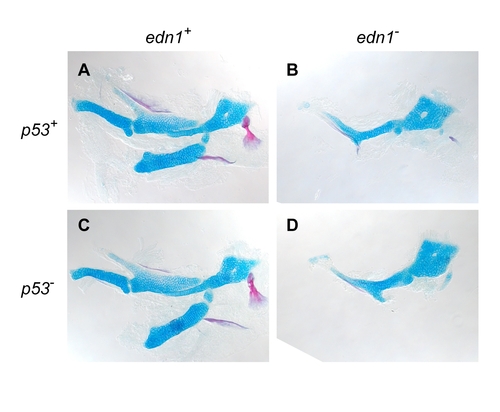
Loss of function of the programmed cell death gene p53 does not rescue the skeletal phenotype of the edn1 mutant. Flat-mount of cartilage and bone stained with Alcian Blue and Alizarin Red. The skeletal phenotypes of the edn1 single mutant and the edn1;p53 double mutant appear identical, whereas phenotypic rescue would be expected if programmed cell death of pharyngeal arch precursor cells accounted for the edn1 hypoplastic skeleton. Hence the experiment argues against cell death as an explanation for the reduced size of the arches. |