
|
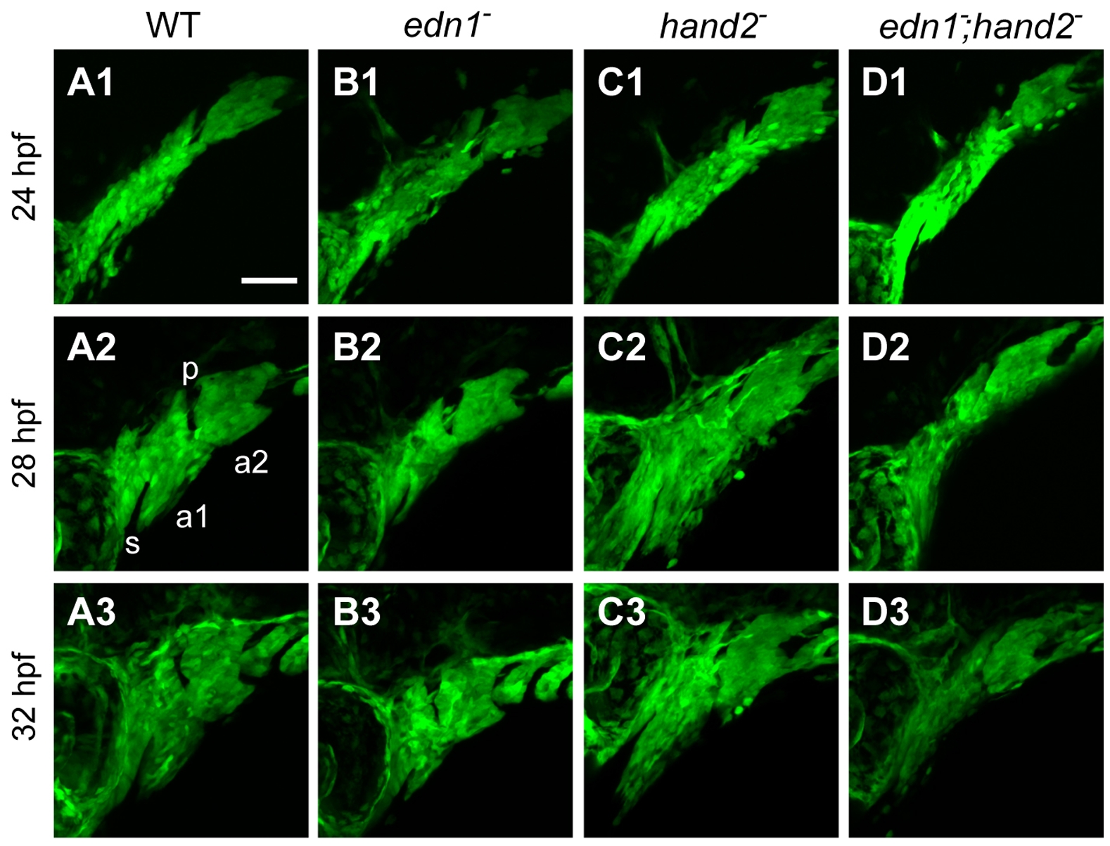
Fig. 2
edn1 and hand2 are required for proper growth and morphogenesis of the early pharyngeal arches.
Projections of lateral confocal images of embryos expressing fli1a:EGFP taken at 24, 28, and 32 hpf. This figure provides an overview of changes in the first two arches (a1, a2, separated by the first pharyngeal pouch, p) that are quantified and more fully documented in subsequent figures. Dorsal is to the top in each panel, and anterior is to the left. No differences in arch morphology are observed among the four genotypes at 24 hpf (upper row of panels). Subsequently the arches prominently lengthen along the DV axis; this DV extension is evidently reduced in the edn1 mutant (B2,B3) and edn1;hand2 double mutant (D2,D3) compared to WT and the hand2 mutant. The arches (particularly the first), also shorten along the AP axis but there appears to be no differences in AP shortening among the genotypes. The hand2 mutant also shows marked expansion of mesenchyme ventral to the stomodeum (s) in the anterior first arch (C2,C3). Scale bar: 50 μm.