
Fig. 1
- ID
- ZDB-FIG-130826-74
- Publication
- Sasaki et al., 2013 - edn1 and hand2 Interact in Early Regulation of Pharyngeal Arch Outgrowth during Zebrafish Development
- Other Figures
- All Figure Page
- Back to All Figure Page
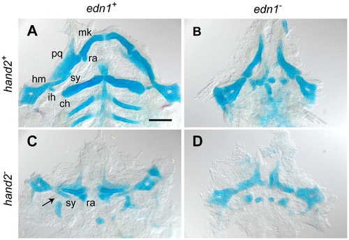
edn1 and hand2 are required for ventral jaw cartilage development. 4 dpf zebrafish skeletons were cartilage and bone stained with Alcian Blue and Alizarin Red, dissected and flat-mounted. (A) WT, with cartilages of the first two arches indicated: In the first arch the dorsal palatoquadrate (pq) and ventral Meckel’s cartilage (mk) articulate, Meckel’s cartilage includes a distinctive retroarticular process (ra) adjacent to the joint. In the second arch the more dorsal hyosymplectic cartilage is subdivided into the hyomandibular (hm) and symplectic (sy) regions, and the prominent ventral cartilage is the ceratohyal (ch). The interhyal cartilage (ih) forms a small hinge within the joint region. (B) In the edn1 larva, ventral cartilages and elements of the joint regions are missing or prominently disrupted. Unidentifiable elements are scattered near the ventral midline. (C) The hand2 larva exhibits ventral reductions similar to edn1, but the arch 2 symplectic cartilage and joint (arrow) are present and the arch 1 retroarticular process is expanded rather than missing. (D) The edn1;hand2 larva exhibits defects similar to edn1; in particular, the symplectic cartilage and retroarticular process cannot be identified. Anterior is upward and right is towards the left. Scale bar: 100 μm. |
| Fish: | |
|---|---|
| Observed In: | |
| Stage: | Day 4 |