
|
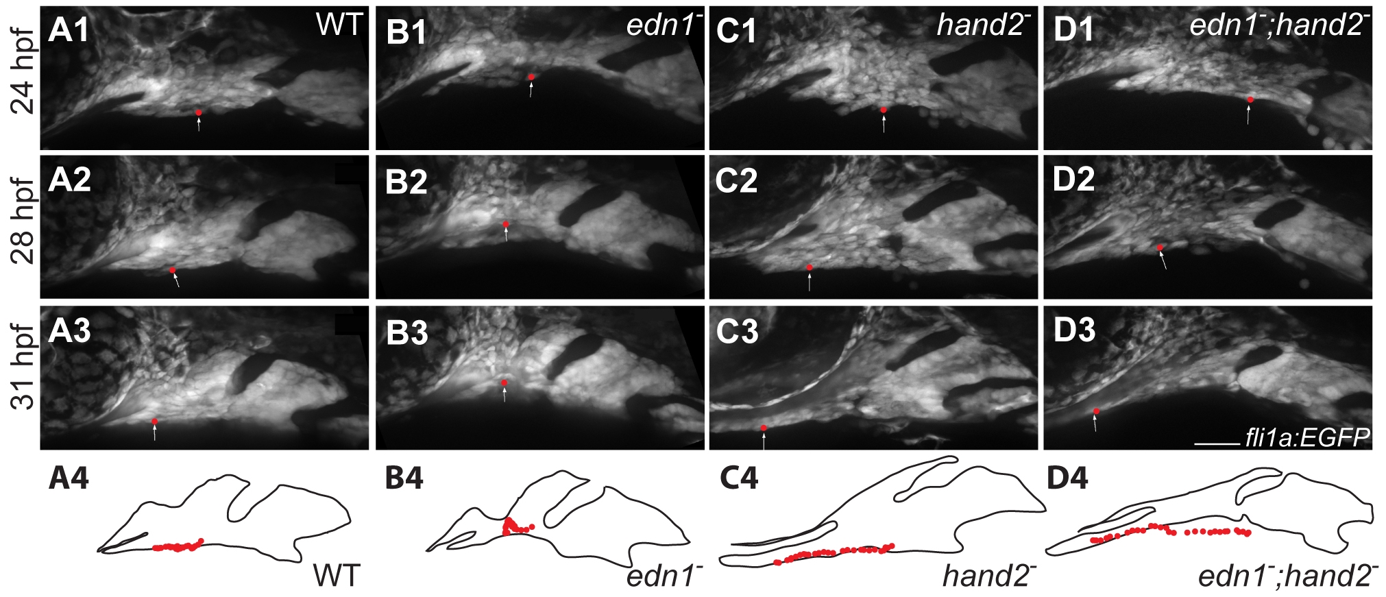
Fig. 7
hand2 negatively regulates anterior flow of ventral arch 1 cells.
fli1a:EGFP animals were imaged by time lapse microscopy from 24–32 hpf. Panels are excerpts from the movie S1 in which maximum confocal projections are presented. Red balls represent the location of an individual cell that was manually tracked in each frame. The positions of tracked cells are overlaid in the outlines. Cells travel from near the midpoint of arch1 to a location posterior to the stomodeum and the eye in wild types and edn1 mutants (A, B). Cells originating at a similar location in hand2 and hand2;edn1 double mutants travel much further, to a position well under the stomodeum and the eye (C, D). Scale bar: 50 μm.