
- Title
-
Comparative gene expression analysis of the fmnl family of formins during zebrafish development and implications for tissue specific functions
- Authors
- Santos-Ledo, A., Jenny, A., and Marlow, F.L.
- Source
- Full text @ Gene Expr. Patterns
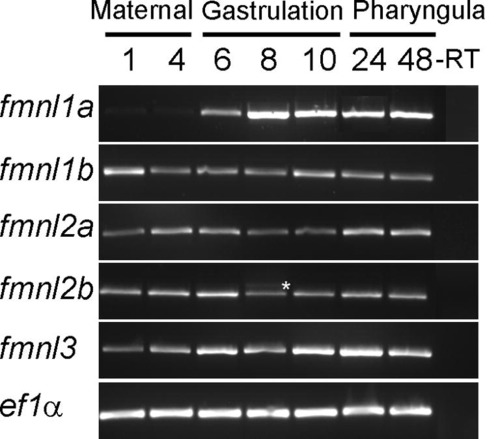
Temporal analysis of zebrafish fmnls using RT-PCR.* indicates alternative splice variant of fmnl2b detected in mid-gastrula stage embryos. This variant includes an additional exon between exons 11 and 12. |
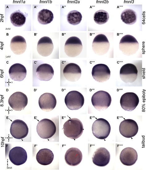
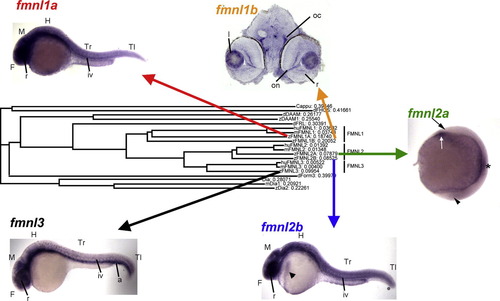
Expression of fmnl transcripts in blastula and gastrula. (A and B) Maternal expression of fmnls (1-4 hpf) and (C-F) expression during gastrulation (5-10 hpf). (E and F) Tailbud stage (Tb). The head is indicated by an arrow and the tailbud by an arrowhead. fmnl2a expression in the otic vesicle primordia (asterisk in E′′), the neuroectoderm border (red arrow in F′′) and in the hatching gland (white arrow in 3E′′). A, anterior; AnVi, anterior view; D, dorsal; DoVi, dorsal view; LaVi, lateral view; P, posterior; V, ventral. |

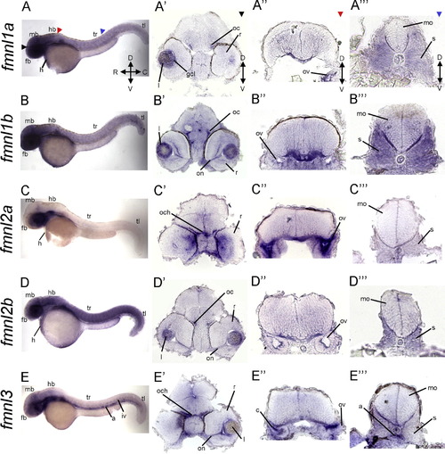
Expression of fmnls at 24 hpf reveals overlapping and distinct patterns. (A-E) Whole mount lateral views. Colored triangles indicate level of cross sections: black indicates forebrain (fb; A′-E′), red at the level of the hindbrain (hb; A′′-E′′), blue denotes sections through the trunk (tr; A′′′-E′′′), and green indicates sections through the tail (tl; a-e). fmnl1a and fmnl1b were detected in the ventral hindbrain in a fibrous-like pattern (arrows in A3 and B3). fmnl1b expression in the dorsal hindbrain (arrowhead in B′′); fmnl2a in the dorsal retina (r) (arrowhead in C′), otic vesicle (ov) (asterisk in C′′) and somites; fmnl2b in the hatching gland (arrowhead in D), the heart (h) and the lens and fmnl3 in the aorta (a), the ov (arrowhead in E′′) and somites (s). Orientation is conserved for all the whole mount and section images. C, caudal; D, dorsal; mb, midbrain; mo, medulla oblongata; n, notochord; iv, intersomitic vessels; R, rostral; V, ventral. EXPRESSION / LABELING:
|
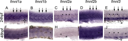
Expression of fmnls in the trunk of larvae at 24 (A-E) and 48 (F-J) hpf. At 24 hpf fmnls were robust in the myotome boundaries (arrows) and fmnl2a and 3 were also expressed in the intersomitic vessels (asterisks) and fmnl3 in the aorta (a) (E). At 48 hpf fmnl1a, 1b and 2b were robust in the notochord (n) (F, G, I) and fmnl3 persisted in the aorta and the intersomitic vessels (asterisks in J). |
Expression of fmnls at 48 hpf reveals more distinct patterns than at earlier stages. (A-E) Lateral views of whole-mount embryos. Colored triangles indicate level of cross sections: black indicates forebrain (fb; A′-E′), red at the level of the hindbrain (hb; A′′-E′′), and blue denotes sections through the trunk (tr; A′′′�E′′′). Colored arrowheads indicate the level cross sections in (A′-E′) black arrows indicate forebrain (fb), (A′′�E′′) at the level of the hindbrain (hb) red arrowhead and (A′′′-E′′′) blue arrowhead indicating sections through the trunk (tr). Enrichment of fmnl1a, 1b and 2b in the head (A, B, D). Robust expression of fmnl1a and 1b in somites (A′′′ and B′′′). fmnl1b, 2b and 3 expression in the visual system including the optic commissure (oc), optic nerve (on) and lens (l) (B′, D′, E′). fmnl2a expression in the medial region of the retina (C′). fmnl2a expression within the otic vesicle (ov), (C′′), while (E′′) Prominent expression of fmnl3 in the cristae (c). a, aorta; C, caudal; gcl, ganglion cell layer; D, dorsal; mb, midbrain; mo, medulla oblongata; n, notochord; och, optic chiasm; r, retina; iv, intersomitic vessels; R, rostral; r, retina; tl, tail; V, ventral. EXPRESSION / LABELING:
|
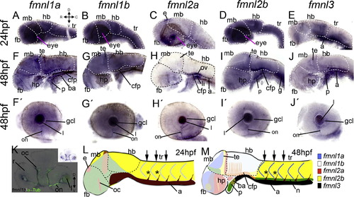
Expression of fmnls in the anterior part of the larvae. At 24 hpf, fmnl1a, 1b and 2b are widely expressed (A, B, D). fmnl2a in the eye and in the epiphysis (e) (C). Enhanced fmnl3 expression in the posterior part of the hindbrain (hb) and in the aorta (a) (E). fmnl1a, 1b and 2b expression in the posterior part of the midbrain (mb) and the floor plate (cfp) of the hb at 48 hpf. fmnl2a and 3 were also detected in the floor plate. and all fmnls were expressed in the optic nerve (on) (F′-J′), this staining colocalized with α-Tubulin (K for fmnl1b). In (L and M), a summary of the expression pattern of all fmnls is shown (not all the domains are represented). ba: branchial arch C, caudal; D, dorsal; fb, forebrain; g: gut; gcl: ganglion cell layer h, heart; hp, hypothalamus; l, lens; li: liver; p: pharynx; R, rostral; r, retina; oc, optic commissure; te: tegmentum; tr, trunk; V, ventral. |
|
Reprinted from Gene expression patterns : GEP, 13(1-2), Santos-Ledo, A., Jenny, A., and Marlow, F.L., Comparative gene expression analysis of the fmnl family of formins during zebrafish development and implications for tissue specific functions, 30-37, Copyright (2013) with permission from Elsevier. Full text @ Gene Expr. Patterns