
Fig. 7
|
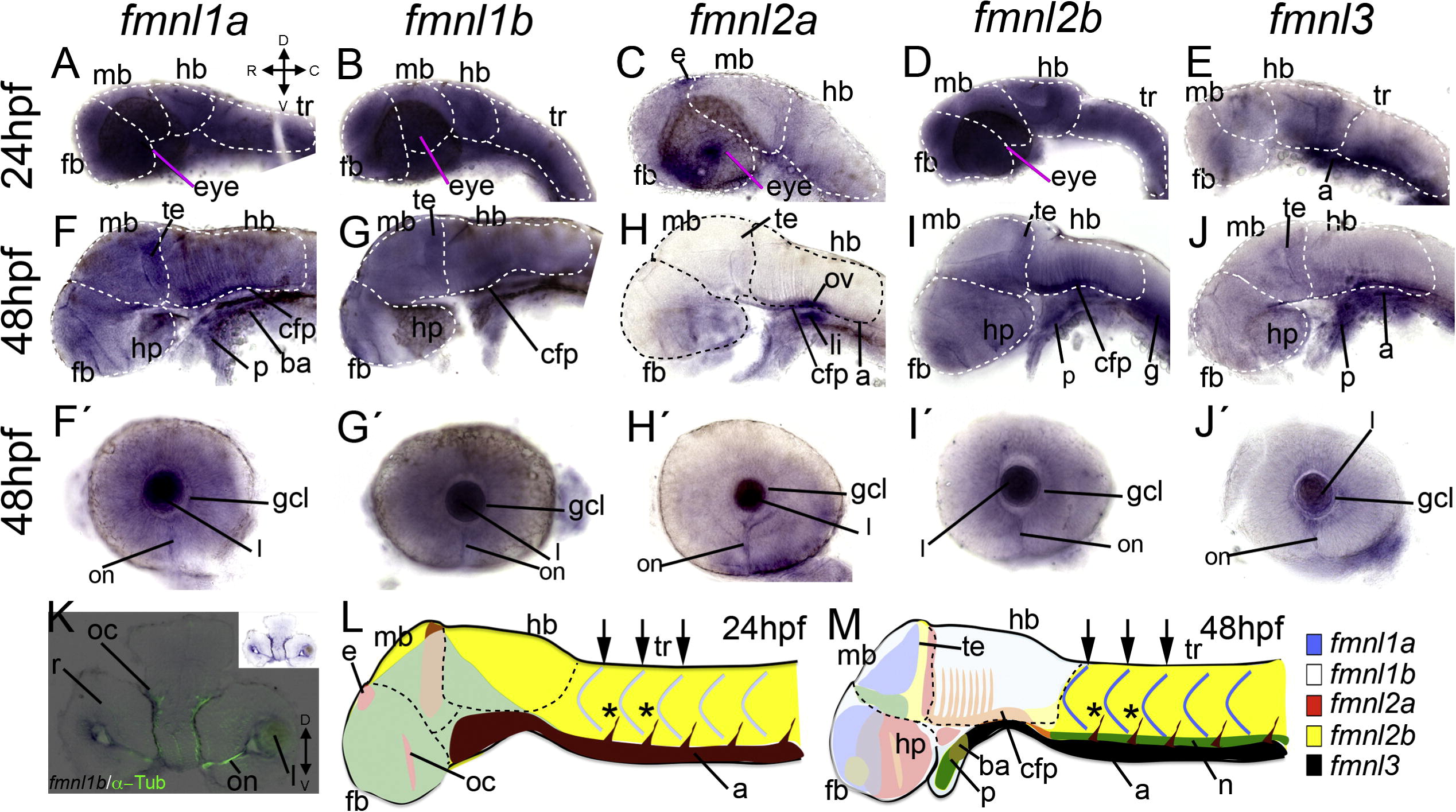
Fig. 7 Expression of fmnls in the anterior part of the larvae. At 24 hpf, fmnl1a, 1b and 2b are widely expressed (A, B, D). fmnl2a in the eye and in the epiphysis (e) (C). Enhanced fmnl3 expression in the posterior part of the hindbrain (hb) and in the aorta (a) (E). fmnl1a, 1b and 2b expression in the posterior part of the midbrain (mb) and the floor plate (cfp) of the hb at 48 hpf. fmnl2a and 3 were also detected in the floor plate. and all fmnls were expressed in the optic nerve (on) (F′-J′), this staining colocalized with α-Tubulin (K for fmnl1b). In (L and M), a summary of the expression pattern of all fmnls is shown (not all the domains are represented). ba: branchial arch C, caudal; D, dorsal; fb, forebrain; g: gut; gcl: ganglion cell layer h, heart; hp, hypothalamus; l, lens; li: liver; p: pharynx; R, rostral; r, retina; oc, optic commissure; te: tegmentum; tr, trunk; V, ventral.
Reprinted from Gene expression patterns : GEP, 13(1-2), Santos-Ledo, A., Jenny, A., and Marlow, F.L., Comparative gene expression analysis of the fmnl family of formins during zebrafish development and implications for tissue specific functions, 30-37, Copyright (2013) with permission from Elsevier. Full text @ Gene Expr. Patterns