
- Title
-
Fluid dynamics in zebrafish Kupffer's vesicle
- Authors
- Okabe, N., Xu, B., and Burdine, R.D.
- Source
- Full text @ Dev. Dyn.
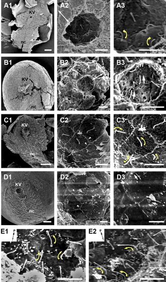
Monocilia project from cells lining both the ventral floor and dorsal roof of Kupffer's vesicle in zebrafish. A-E: Scanning electron microscopy (SEM) analysis of Kupffer's vesicle at the 3-somite stage (A,B), 7-somite stage (E1), 9-somite stage (E2), and 11- to 12-somite stage (C,D). In each row of A-D, higher magnification images are presented with low magnification in the left to highest magnification at the right. Monocilia (arrows) project from individual cells. Cells are distinguishable due to their cell borders (arrowheads). Each image is representative of embryos at these stages (Nemb = 5 for 3- to 4-somite stages, Nemb = 6 for 7-somite stages, Nemb = 11 for 9-somite stages, Nemb = 11 for 11 somite stages). Double-headed arrows indicate cells viewed laterally. Cilia direct toward the posterior (B3,D3, white arrows) and the cilia at the dorsal-anterior roof (A3, C3, E1, E2) bend toward the membrane (yellow arrows). In all images, posterior is to the top. Inset in E1, E2 denotes the posterior of the embryo. The left side of the embryo is to the right side of the panels in B1-B3, D1-D3 and to the left side of the panels in A1-A3, C1-C3, E1-E2. Scale bars = 50 μm in A1,B1,C1,D1, 5 μm in A2-A3,B2-B3,C2-C3,D2-D3,E1-E2. |
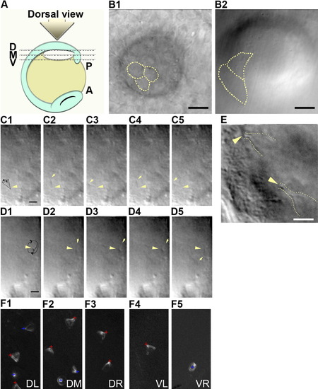
Cilia rotation at different levels within Kupffer's vesicle. A: Schematic diagram for taking movies within zebrafish Kupffer's vesicle. The dorsal roof of Kupffer's vesicle faces up (dorsal view). We focused the lens to either the dorsal (line D), middle (line M), or ventral (line V). B: Single differential interference contrast (DIC) microscopy image showing morphological features; at the dorsal roof (B1) and the ventral floor (B2) at 6-somite stage. Dotted lines mark cell-cell contacts. C,D: Cilia at the 3-somite stage appear to rotate counterclockwise at the dorsal roof (C1-C5), and clockwise at the ventral floor (D1-D5). The trajectories of cilia motility on dorsal roof (C1) and on the ventral floor (D1) are traced with a black line. Arrows mark the top of the cilium; arrowheads mark the basal body of the cilium. E: Combination of four stills of the movies taken at anterior-dorsal roof showed the conical shape of cilia movement (dotted line) at 9-somite stage. Yellow arrowheads indicate the basal bodies of the cilia. F: Sum of the trajectories of cilia motility at 7- to 8-somite stages. The dorsal roof (F1-F3) and the ventral floor regions (F4-F5), which are relatively flat within the vesicle. Red cross; basal body of cilium that points toward the posterior, Blue cross; basal body of cilium that is not pointing toward the posterior. Posterior side of the embryo is to the right in (E), to down in (F). DL, dorsal-left; DM, dorsal-middle; DR, dorsal right; VL, ventral-left; VR, ventral-right. Scale bars = 8 μm in B1-B2, 3.2 μm in C1-C5,D1-D5, 4 μm in E. |
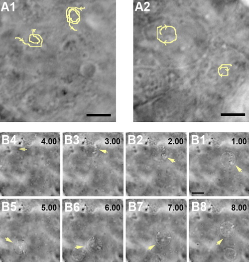
Specification of local flow around individual cilium and dominant net flow inside Kupffer's vesicle. A1,A2: Counterclockwise movements of beads closest to the dorsal roof (A1), and clockwise movements of the beads closest to the ventral floor (A2) at 5-somite stage. Trajectories of beads movements are indicated in yellow. B1-B8: The dominant counterclockwise flow in the middle of the vesicle observed by the movement of a bubble (arrows) at 9-somite stage. There is a 1-sec interval between stills. Posterior side of the embryo is to the right in (B). Scale bars = 4 μm in A1-A2, 8 μm in B1-B8. |
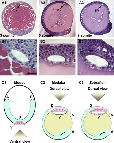
Histological analysis shows more cells at the anterior and dorsal roof of Kupffer's vesicle. Lateral sections of Kupffer's vesicle stained by hematoxylin and eosin (H&E staining) at different stages (Nemb = 5 for each stage). A,B: Lateral views of zebrafish embryos were obtained by sectioning JB-4 resin at 3-somite (A1,B1), at 5-somite (A2,B2), and at 9-somite stages (A3,B3). In all panels, the dorsal side of Kupffer's vesicle (arrows) is up, with the posterior of the embryo to the top of the panel. All cells were assumed to locate to the luminal surface of Kupffer's vesicle. Brackets denote the anterior-dorsal regions of Kupffer's vesicle where cells are more numerous. Scale bar = 100 μm in (A1-A3), 20 μm in (B1-B3). C: Schematic diagrams of mouse node (C1), medaka Kupffer's vesicle (C2), and zebrafish Kupffer's vesicle (C3). The ciliated cells are shown in pink with blue nuclei and green cilia. Fish yolk is labeled in yellow. Note that the medaka and zebrafish embryos are positioned upside down from how they are normally presented such that the dorsal roof of Kupffer's vesicle is in the same orientation as the ventral mouse node. |