
Image
|
Figure Caption
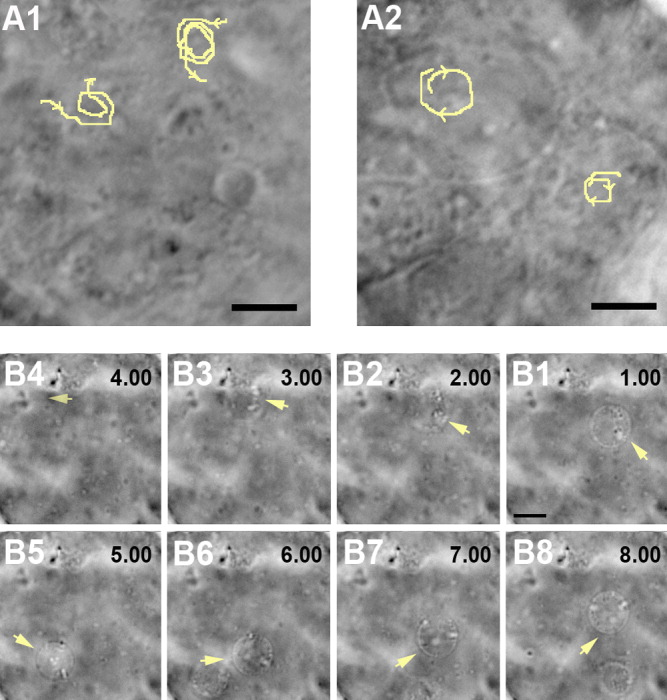
Fig. 4 Specification of local flow around individual cilium and dominant net flow inside Kupffer's vesicle. A1,A2: Counterclockwise movements of beads closest to the dorsal roof (A1), and clockwise movements of the beads closest to the ventral floor (A2) at 5-somite stage. Trajectories of beads movements are indicated in yellow. B1-B8: The dominant counterclockwise flow in the middle of the vesicle observed by the movement of a bubble (arrows) at 9-somite stage. There is a 1-sec interval between stills. Posterior side of the embryo is to the right in (B). Scale bars = 4 μm in A1-A2, 8 μm in B1-B8.
Acknowledgments
This image is the copyrighted work of the attributed author or publisher, and
ZFIN has permission only to display this image to its users.
Additional permissions should be obtained from the applicable author or publisher of the image.
Full text @ Dev. Dyn.