
|
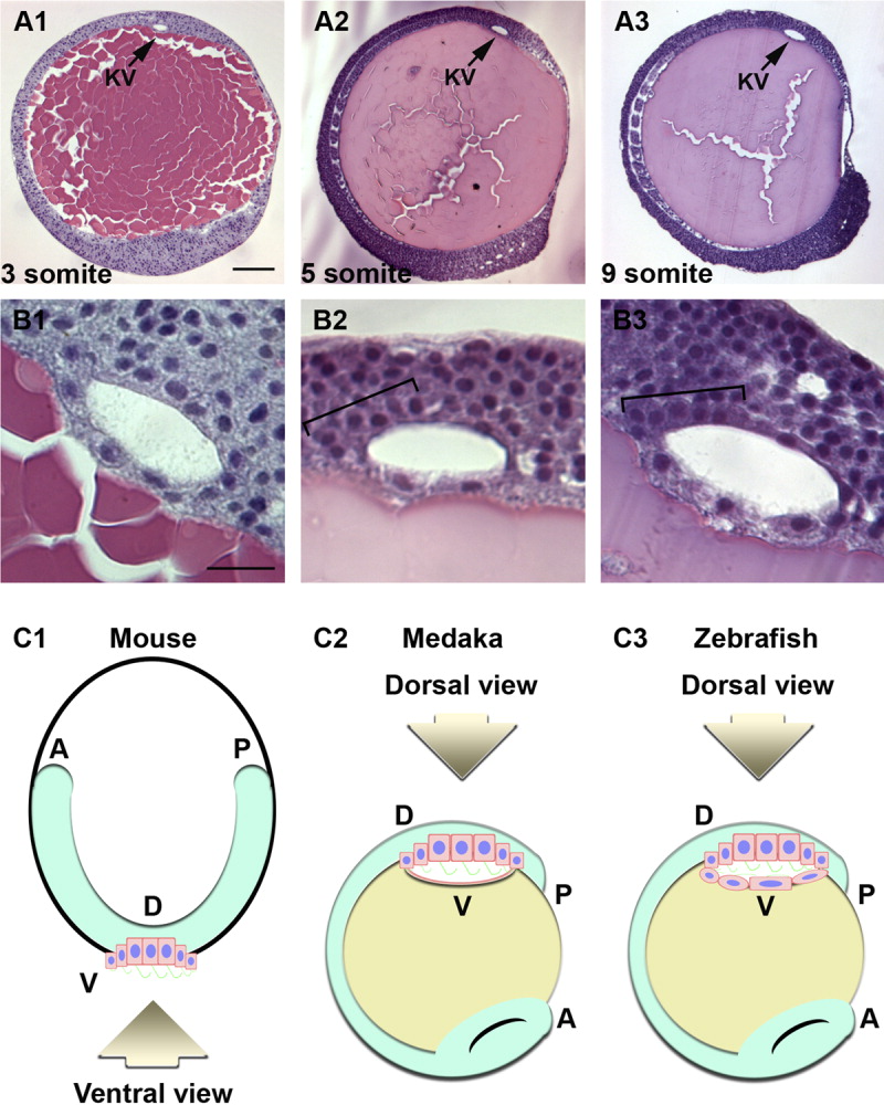
Fig. 5 Histological analysis shows more cells at the anterior and dorsal roof of Kupffer's vesicle. Lateral sections of Kupffer's vesicle stained by hematoxylin and eosin (H&E staining) at different stages (Nemb = 5 for each stage). A,B: Lateral views of zebrafish embryos were obtained by sectioning JB-4 resin at 3-somite (A1,B1), at 5-somite (A2,B2), and at 9-somite stages (A3,B3). In all panels, the dorsal side of Kupffer's vesicle (arrows) is up, with the posterior of the embryo to the top of the panel. All cells were assumed to locate to the luminal surface of Kupffer's vesicle. Brackets denote the anterior-dorsal regions of Kupffer's vesicle where cells are more numerous. Scale bar = 100 μm in (A1-A3), 20 μm in (B1-B3). C: Schematic diagrams of mouse node (C1), medaka Kupffer's vesicle (C2), and zebrafish Kupffer's vesicle (C3). The ciliated cells are shown in pink with blue nuclei and green cilia. Fish yolk is labeled in yellow. Note that the medaka and zebrafish embryos are positioned upside down from how they are normally presented such that the dorsal roof of Kupffer's vesicle is in the same orientation as the ventral mouse node.