
|
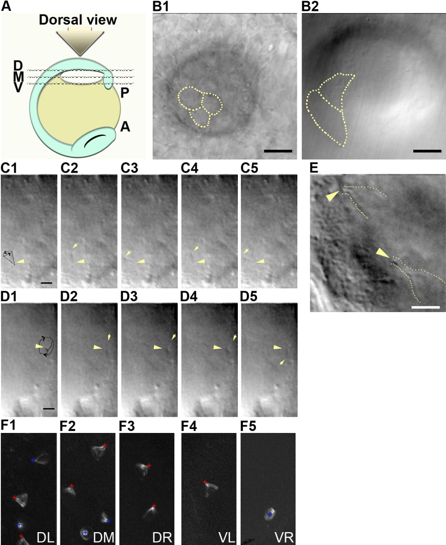
Fig. 3 Cilia rotation at different levels within Kupffer's vesicle. A: Schematic diagram for taking movies within zebrafish Kupffer's vesicle. The dorsal roof of Kupffer's vesicle faces up (dorsal view). We focused the lens to either the dorsal (line D), middle (line M), or ventral (line V). B: Single differential interference contrast (DIC) microscopy image showing morphological features; at the dorsal roof (B1) and the ventral floor (B2) at 6-somite stage. Dotted lines mark cell-cell contacts. C,D: Cilia at the 3-somite stage appear to rotate counterclockwise at the dorsal roof (C1-C5), and clockwise at the ventral floor (D1-D5). The trajectories of cilia motility on dorsal roof (C1) and on the ventral floor (D1) are traced with a black line. Arrows mark the top of the cilium; arrowheads mark the basal body of the cilium. E: Combination of four stills of the movies taken at anterior-dorsal roof showed the conical shape of cilia movement (dotted line) at 9-somite stage. Yellow arrowheads indicate the basal bodies of the cilia. F: Sum of the trajectories of cilia motility at 7- to 8-somite stages. The dorsal roof (F1-F3) and the ventral floor regions (F4-F5), which are relatively flat within the vesicle. Red cross; basal body of cilium that points toward the posterior, Blue cross; basal body of cilium that is not pointing toward the posterior. Posterior side of the embryo is to the right in (E), to down in (F). DL, dorsal-left; DM, dorsal-middle; DR, dorsal right; VL, ventral-left; VR, ventral-right. Scale bars = 8 μm in B1-B2, 3.2 μm in C1-C5,D1-D5, 4 μm in E.