FIGURE SUMMARY
Title

Shp2 Knockdown and Noonan/LEOPARD Mutant Shp2-Induced Gastrulation Defects

Authors
Jopling, C., van Geemen, D., and den Hertog, J.
Source
Full text @ PLoS Genet.

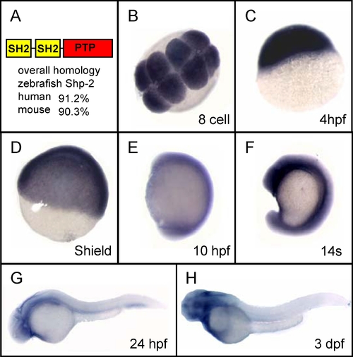
Zebrafish Shp2 Is Conserved and Is Expressed Ubiquitously during Development
(A) Schematic representation of zebrafish Shp2 with two SH2 domains to the N-terminal side of the PTP domain. The overall sequence identity with human and mouse Shp2 is indicated. (B–F) In situ hybridization with a Shp2-specific antisense probe at various stages of development: (B) 8-cell stage, (C) 4 hpf, (D) shield stage, (E) 10 hpf, (F) 14 somite (14 s, 14 hpf), (G) 24 hpf, and (H) 3 dpf.

EXPRESSION / LABELING:
Gene:
Fish:
Anatomical Terms:
Stage Range: 8-cell to Protruding-mouth

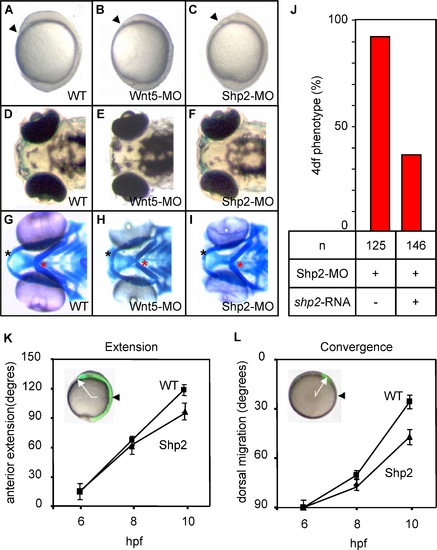
Shp2-MO–Induced CE Cell Movement Defects
Zebrafish embryos were not injected (A, D, G) or microinjected with Wnt5-MO (5 ng) (B, E, H) or Shp2-MO (1.0 ng) (C, F, I) at the one-cell stage.
(A–C) Morphology at 10 hpf shows reduced anterior extension of the Wnt5-MO– and Shp2-MO–injected embryos. Arrowheads indicate the anterior of the embryos
(D–F) Morphology of the Wnt5 and Shp2 knockdown embryos at 4 dpf show a mild hammerhead-like phenotype.
(G–I) Alcian blue staining of the cartilage in the heads of 4 dpf embryos. Black asterisk, Meckel′s cartilage; red asterisk, ceratohyal. (J) Zebrafish embryos were (co-) injected with Shp2-MO (1.0 ng) and 300 pg human shp2 mRNA and scored at 4 dpf.
(K,L) Embryos were loaded with caged fluorescein dextran and the fluorophore was uncaged at the shield stage (6 hpf) dorsally to determine anterior extension (K, white arrow in inset; site of uncaging, black arrowhead) or laterally to determine dorsal migration (L, white arrow in inset; initial position at the shield stage, black arrowhead). Cell labeling of the same embryos was followed immediately after uncaging at 80% epiboly (8 hpf) and at tailbud stage (10–10.5 hpf). WT and Shp2-MO–injected embryos were assessed and averages for ten embryos are given in degrees.

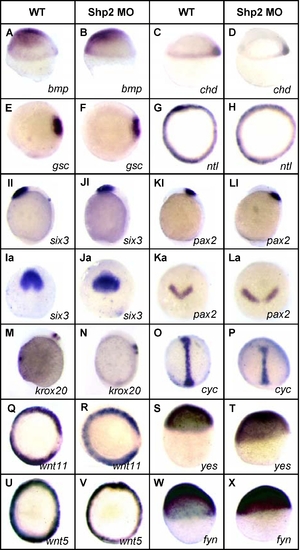
Shp2 Knockdown Did Not Affect Cell Specification
Control and Shp2-MO–injected embryos were fixed at 6 hpf (A–H, Q–X), 8 hpf (O,P), or 10 hpf (I–L), and in situ hybridization was done with the indicated probes. Either lateral views (A–D, Il–Ll, M, N, S, T, W, X) or animal pole views (E–H, Ia-La, O–R, U, V) are depicted here.

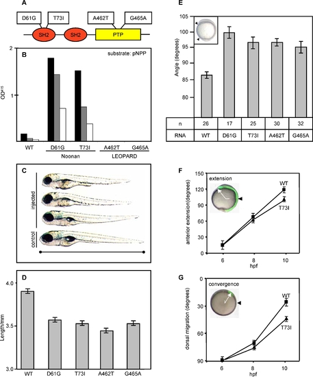
NS and LS Mutant Shp2 Expression Induced CE Cell Movement Defects during Gastrulation
(A) Schematic representation of zebrafish Shp2 with the NS and LS mutations indicated.
(B) PTP activity of WT Shp2, NS (D61G and T73I) and LS (A462T and G465A) mutant Shp2 was assayed using p-nitrophenylphosphate and quantified spectrophotometrically. Each experiment was done with three different amounts of GST-fusion protein (black bar, high; grey bar, middle; and white bar, low).
(C) NS- and LS-Shp2 expression reduced zebrafish embryo body length. Three injected embryos (D61G, 150pg) at 4 dpf are depicted with a noninjected control embryo at the bottom. The figure is representative of defects associated with all NS- and LS-Shp2 expressing embryos.
(D) The length of the embryos was measured at 4 dpf and the average is shown here. Two tailed student t-tests indicate a significant decrease in length after injection of each NS or LS RNA ( p < 0.001).
(E) NS- and LS-Shp2 RNA injection results in reduced extension of the body axis at 10 hpf. The angle between the most anterior and posterior embryonic structure (arrowheads in inset) was determined at the one-somite stage and the average angle is depicted here in degrees. Two tailed student t-tests indicate a significant increase in the angle after injection of NS or LS RNA (p < 0.001). The number of embryos used here is indicated (n).
(F, G) Cell tracing was done as described in Figure 2. NS-Shp2 (T73I) was coinjected with caged fluorophore at the one-cell stage and after uncaging at 6 hpf, extension and convergence were determined at 8 hpf and 10 hpf in ten embryos per condition.

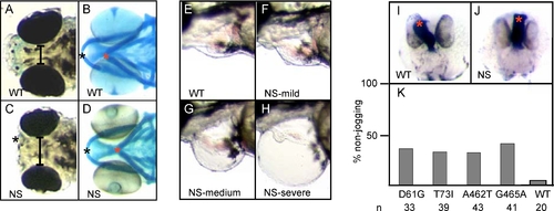
Craniofacial and Heart Defects upon NS or LS RNA Injection
(A,C) Extension of anterior structures (black asterisk) was impaired and the eyes were spaced wider apart (black bar) in NS-Shp2–injected embryos (D61G, 150 pg) (C) than in the noninjected control (A). The figure is representative of defects associated with all NS- and LS-Shp2–injected embryos. (B,D) Alcian blue staining of the cartilage in the head of NS-Shp2 injected embryos (D) compared to noninjected control (B). Black asterisk, Meckel's cartilage; red asterisk, ceratohyal. (E–K) NS- and LS-induced heart defects. (E–H) Morphology of representative 3 dpf embryos with (E) noninjected control, (F) mild, (G) intermediate, and (H) severe heart defect. Phenotypes evoked by NS- and LS-Shp2 were indistinguishable. (I–K) cmlc2 in situ hybridization marks the heart. (I) Noninjected control with normal heart jogging (red asterisk). (J) Injected embryo showing defective cardiac jogging (red asterisk). (K) Quantification of heart jogging of NS- or LS-Shp2 RNA–injected embryos stained with cmlc2, depicted as percent non-jogging.

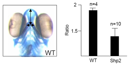
Morphometry of the Hammerhead Phenotype
The distance between the eyes and the distance from the middle of the eyes to the tip of the nose (as indicated in the left panel) was determined in 4 dpf embryos after staining with alcian blue (see Materials and Methods). The ratio of the distance to the tip of the nose and the distance between the eyes was determined and is plotted in the bar graph, depicted in the panel on the right. Four WT and ten Shp2-MO injected embryos were analyzed. There is a significant difference in ratios between the WT (1.87 ± 0.03) and Shp2 knockdown (1.40 ± 0.20), indicating that Shp2 knockdown indeed induced a hammerhead phenotype.

PHENOTYPE:
Fish:
Knockdown Reagent:
Observed In:
Stage: Day 4

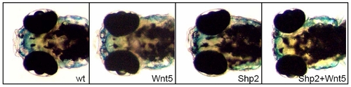
Shp2 and Wnt5 Knockdown Acts Synergistically to Induce a Hammerhead Phenotype at 4 dpf
Shp2-MO and Wnt5-MO were injected at 50% of their normal concentration. By themselves, these injections did not induce defects (compare to morphology at 4 dpf with WT, noninjected control). Coinjection of Shp2-MO and Wnt5-MO did induce the hammerhead phenotype at 4 dpf. Note the blunted face and the increased distance between the eyes in the double-injected embryo (Shp2 + Wnt5).

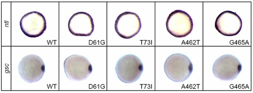
Expression of NS- and LS-Shp2 Did Not Induce Defects in Cell Specification
Synthetic RNA encoding NS-Shp2 (D61G or T73I) or LS-Shp2 (A462T or G465A) was injected into one-cell stage zebrafish embryos. The embryos were fixed at 6 hpf and in situ hybridization was done with the indicated probes. Animal pole views are depicted here. Expression of the mesendodermal marker ntl and of the organiser marker gsc remained unchanged in the NS- and LS-Shp2–expressing embryos.

Acknowledgments
This image is the copyrighted work of the attributed author or publisher, and ZFIN has permission only to display this image to its users. Additional permissions should be obtained from the applicable author or publisher of the image. Full text @ PLoS Genet.