
- Title
-
Slit1a inhibits retinal ganglion cell arborization and synaptogenesis via Robo2-dependent and -independent pathways
- Authors
- Campbell, D.S., Stringham, S.A., Timm, A., Xiao, T., Law, M.Y., Baier, H., Nonet, M.L., and Chien, C.B.
- Source
- Full text @ Neuron
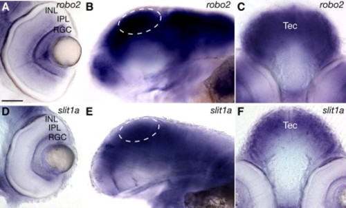
Zebrafish slit1a and robo2 Are Expressed in the Eye and Optic Tectum during RGC Arborization and Synaptogenesis Shown are whole-mount lateral views ([B and E]; anterior left, dorsal up), and transverse vibratome sections through the retina ([A and D]; dorsal up) and optic tectum ([C and F]; dorsal up) at 76 hpf. robo2 is highly expressed throughout the tectum and RGCs (A–C). slit1a is strongly expressed throughout the tectum and weakly in RGCs (D–F). RGC, retinal ganglion cells; IPL, inner plexiform layer; INL, inner nuclear layer. Dashed ovals indicate the tectum. Scale bar, 50 μm. EXPRESSION / LABELING:
|
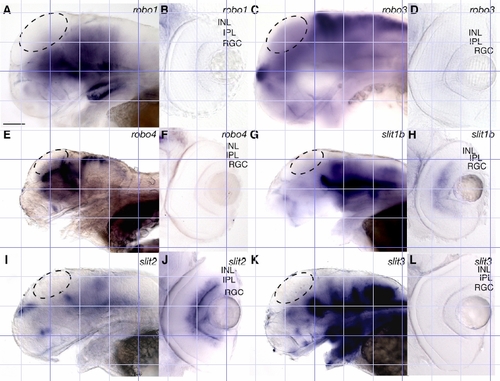
Expression patterns of slit and robo family members during arborisation and synaptogenesis. Shown are whole-mount lateral views (A, C, E, G, I and K; anterior left, dorsal up) and vibratome sections through the retina (B, D, F, H, J and L; dorsal up) at 76 hpf. robo1 and robo4 are not expressed in the tectum or RGCs (A-B, E-F). robo3 is not expressed widely in the tectum (C) but is expressed in a few discrete tectal cells outside the plane of focus. robo3 is not expressed in RGCs (D). slit1b, slit2, and slit3 are not detectably expressed in the tectum (G, I, K). slit1b is expressed strongly in the inner nuclear layer (H), slit2 is expressed strongly in the inner plexiform layer (J) and slit3 is not expressed in the retina (L). RGC, retinal ganglion cells; IPL, inner plexiform layer; INL, inner nuclear layer. Dashed ovals indicate the tectum. Scale bar = 50μm. EXPRESSION / LABELING:
|
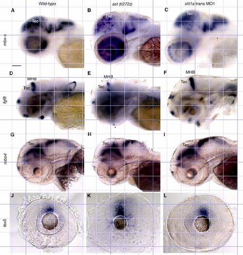
Gross brain and eye patterning are unaffected in ast and slit1a morphant embryos. In situ hybridisation of 76 hpf WT, ast (ti272z) and slit1a morphant embryos with antisense mRNA probes for mbx-s (A-C), fgf8 (D-F), robo4 (G-I) and tbx5 (J-L). No changes in the expression patterns of these genes were observed between WT, ast (ti272z) or slit1a morphant embryos. Tec, tectum; MHB, midbrain-hindbrain boundary. Lateral views of wholemounts (A-I). Eyes only (J-L). Anterior left, dorsal up. Scale bar = 100μm. Supplemental EXPRESSION / LABELING:
|