
- Title
-
TALE-Family homeodomain proteins regulate endodermal sonic hedgehog expression and pattern the anterior endoderm
- Authors
- diIorio, P., Alexa, K., Choe, S.K., Etheridge, L., and Sagerström, C.G.
- Source
- Full text @ Dev. Biol.

meis3 is expressed in the endoderm anterior to the developing islet. (A–C) 24 hpf zebrafish embryos labeled by double in situ hybridization (both probes detected in purple) for insulin (black arrows) together with meis1 (A), meis2 (B) or meis4 (C) reveal that these meis genes are not expressed in the anterior endoderm. Both meis1 and meis2 are expressed in the neural tube (white asterisks). (D, E) At 24 hpf, meis3 (purple stain; black arrow) is expressed in endoderm just anterior to the insulin-expressing islet (red stain; red arrow). (F–H) Expression of meis1, meis2 and meis4 cannot be detected in the gut at 48 hpf (embryos co-labeled for insulin expression [black arrow]; both probes detected in purple). (I) meis3 (purple stain; black arrow) is expressed adjacent to the insulin-expressing islet (red stain, red arrow) at 48 hpf. (J) At 48 hpf, pdx1 expression is seen in duodenal cells and putative duct progenitors as well as the islet (yellow outline). (K) shh (purple stain; black arrow) is expressed adjacent to the insulin-expressing islet (red stain; red arrow) at 48 hpf . Developmental stage is indicated at lower left, probes are indicated at lower right. Lateral views in panels A–D; dorsal views in panels E, H–K; ventrolateral views in panels F, G. Anterior is to the left in all panels. |
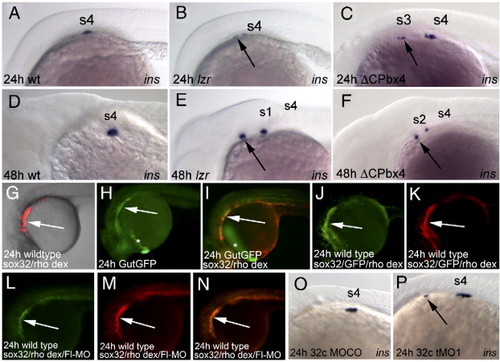
meis3 and pbx4 are required to prevent insulin expression in the anterior endoderm. (A–F) Ectopic insulin-expressing cells arise in the anterior endoderm of meis3 and pbx4-deficient embryos. Wild type embryos (A, D), lzr mutant embryos (B, E) and embryos injected with the dominant negative Meis construct (ΔCPbx4; C, F) were assayed for insulin expression at 24 hpf (A–C) and 48 hpf (D–F). Ectopic insulin expression is indicated by arrows. Note that all insulin positive cells become shifted anteriorly by 48 hpf (see text for details). (G–I) Cells injected with sox32 mRNA at the 32-cell stage primarily populate the endoderm. Wild type (G) or GutGFP transgenic (H, I; (Field et al., 2003)) embryos were injected with sox32 mRNA and rhodamine dextran at the 32 cell stage. At 24 hpf, the rhodamine signal is observed primarily in a deep layer of the embryo adjacent to the yolk (panel G shows a confocal stack of ∼125 μm that extends about halfway through the embryo). This region corresponds to the endoderm, since the rhodamine signal coincides with the GFP-expressing endoderm (white arrows in panels H, I), but not the GFP-expressing hatching gland (white asterisks in panels H, I) of GutGFP embryos. White arrows point to endoderm. (J, K) mRNA and rhodamine dextran co-localize in the endoderm following co-injection at the 32-cell stage. Wild type embryos were injected with sox32 mRNA, GFP mRNA and rhodamine dextran at the 32-cell stage and monitored for GFP (J) and rhodamine (K) expression at 24 hpf. White arrows point to the endoderm. (L–N) MOs co-localize with the rhodamine lineage label in endoderm following co-injection at the 32-cell stage. Wild type embryos were injected with sox32 mRNA, rhodamine dextran and fluorescein-tagged MO at the 32-cell stage. Embryos were raised to 24 hpf and monitored for fluorescein (L) and rhodamine (M) expression. Panel N is an overlay of panels L and M. White arrows point to endoderm. (O, P) Targeting of a meis3 MO (tMO1) to the endoderm produces ectopic anterior insulin expression (arrow in panel P). All panels are lateral views with anterior to the left. s1–s4 denotes the position of somites 1–4 as determined using Nomarski optics. |
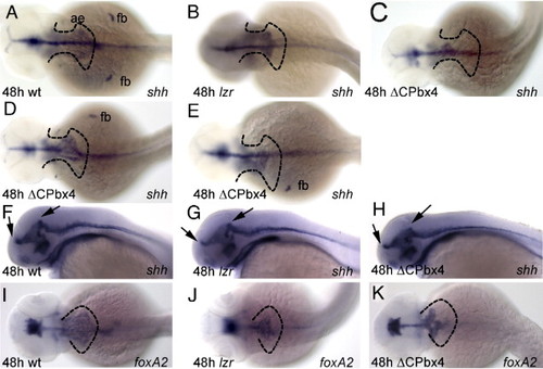
shh and foxa2 expression is disrupted in the anterior endoderm of pbx4- and meis-deficient embryos. (A) shh is expressed in the anterior endoderm (ae) and fin buds (fb) of 48 hpf wild type embryos. (B) lzr mutants (as identified by the absence of pectoral fin buds) exhibit reduced shh expression in the anterior endoderm. (C–E) shh expression is reduced in the anterior endoderm and fin buds of embryos injected with the dominant negative Meis construct (ΔCPbx4). Variations in the severity of the ΔCPbx4-mediated phenotype are likely due variable distribution of the injected mRNA, as routinely seen with this type of injections. (F–H) Prominent sites of neurectodermal shh expression (black arrows) are unaltered in lzr mutant and meis-deficient embryos. (I) foxa2 is expressed in the anterior endoderm of 48 hpf embryos. (J, K) Disruption of foxa2 expression in lzr mutant and meis-deficient embryos is similar to that seen for shh. Dashed black lines outline the normal expression domains of shh (A-E) and foxa2 (I-K) in the endoderm. Dorsal views in panels A-E, I-K; lateral views in panels F-H. Anterior is to the left in all panels. |
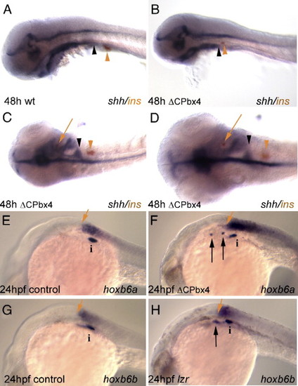
Ectopic insulin expression in meis-deficient embryos coincides with loss of shh expression. (A) insulin expressing cells (orange arrowhead) develop just caudal to the posterior margin of pharyngeal shh expression (black arrowhead). (B, C) This spatial relationship is conserved in meis-deficient embryos exhibiting caudally reduced shh expression (compare the position of arrowheads in panel A with the position in panels B, C). (D) Higher magnification of meis-deficient embryo in panel C demonstrating the appearance of insulin expressing cells (red arrow) in a shh negative region of the anterior endoderm. (E–H) hox gene expression in mesoderm is unaffected in meis-deficient embryos and lzr mutants. Mesodermal expression of hoxb6a (E) and hoxb6b (G) normally terminates just anterior to the principal islet (i). Ectopic insulin expressing cells develop anterior to the rostral limits of hox expression in meis-deficient (F) and lzr (H) embryos. In both classes of embryos the anterior limit of hox expression is unchanged relative to the principal islet. Panels A, B, E–H are in lateral view; panels C and D are in dorsal view. Anterior is to the left in all panels. i, islet; black arrow indicates ectopic insulin-expressing cells; red arrows in panels E–H indicate anterior limit of hox gene expression. |
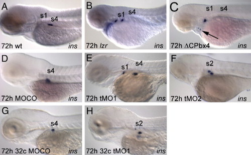
Disruption of meis3 and pbx4 function causes an anterior shift of insulin-positive cells. Control (A), lzr mutant (B), δCPbx4-injected (C), MOCO-injected (D), tMO1-injected (E) and tMO2-injected (F) embryos, as well as embryos where MOCO (G) or tMO1 (H) was targeted to the endoderm, were assayed for insulin expression by in situ hybridization at 72 hpf. Note that all insulin positive cells are found anterior to somite 4 in experimental embryos (B, C, E, F, H), but not in control embryos (A, D, G). s1–s4 indicate the position of somites 1–4. All embryos are in lateral view with anterior to the left. EXPRESSION / LABELING:
PHENOTYPE:
|
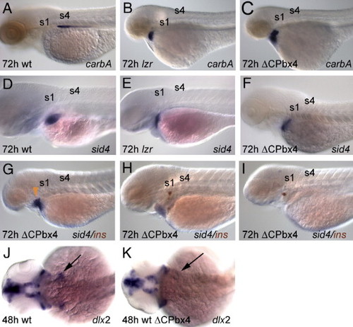
Multiple organs are displaced anteriorly in meis- and pbx4-deficient embryos. 72 hpf lzr mutant and meis-deficient embryos exhibit anterior displacement of exocrine pancreas (carboxypeptidase A, A–C) and liver (sid4, D–F) gene expression. (G–I) Anterior shifts in meis-deficient embryos can result in loss of liver-, but not islet-specific gene expression. (J, K) Posterior, dlx2-expressing arches are absent from meis-deficient embryos. Panels A–I are in lateral view; panels J, K are in dorsal view. Anterior is to the left in all panels. EXPRESSION / LABELING:
PHENOTYPE:
|
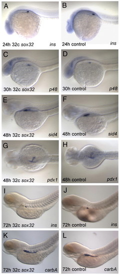
sox32 mRNA does not perturb normal endoderm development. A single cell was injected with sox32 mRNA and rhodamine dextran at the 32-cell stage as outlined in Materials and methods (A, C, E, G, I, K), or left uninjected (B, D, F, H, J, L). Embryos were fixed and assayed for expression of insulin at 24 hpf (A, B), p48 at 30 hpf (C, D), sid4 at 48 hpf (E, F), pdx1 at 48 hpf (G, H), insulin at 72 hpf (I, J) and carbA at 72 hpf (K, L) by whole mount in situ hybridization. All embryos are in lateral view with anterior to the left. EXPRESSION / LABELING:
|
Reprinted from Developmental Biology, 304(1), diIorio, P., Alexa, K., Choe, S.K., Etheridge, L., and Sagerström, C.G., TALE-Family homeodomain proteins regulate endodermal sonic hedgehog expression and pattern the anterior endoderm, 221-231, Copyright (2007) with permission from Elsevier. Full text @ Dev. Biol.