
|
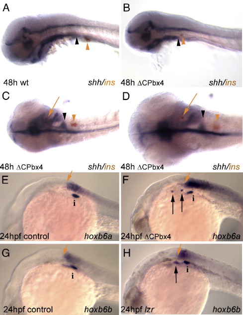
Fig. 4 Ectopic insulin expression in meis-deficient embryos coincides with loss of shh expression. (A) insulin expressing cells (orange arrowhead) develop just caudal to the posterior margin of pharyngeal shh expression (black arrowhead). (B, C) This spatial relationship is conserved in meis-deficient embryos exhibiting caudally reduced shh expression (compare the position of arrowheads in panel A with the position in panels B, C). (D) Higher magnification of meis-deficient embryo in panel C demonstrating the appearance of insulin expressing cells (red arrow) in a shh negative region of the anterior endoderm. (E–H) hox gene expression in mesoderm is unaffected in meis-deficient embryos and lzr mutants. Mesodermal expression of hoxb6a (E) and hoxb6b (G) normally terminates just anterior to the principal islet (i). Ectopic insulin expressing cells develop anterior to the rostral limits of hox expression in meis-deficient (F) and lzr (H) embryos. In both classes of embryos the anterior limit of hox expression is unchanged relative to the principal islet. Panels A, B, E–H are in lateral view; panels C and D are in dorsal view. Anterior is to the left in all panels. i, islet; black arrow indicates ectopic insulin-expressing cells; red arrows in panels E–H indicate anterior limit of hox gene expression.
Reprinted from Developmental Biology, 304(1), diIorio, P., Alexa, K., Choe, S.K., Etheridge, L., and Sagerström, C.G., TALE-Family homeodomain proteins regulate endodermal sonic hedgehog expression and pattern the anterior endoderm, 221-231, Copyright (2007) with permission from Elsevier. Full text @ Dev. Biol.