
- Title
-
Depletion of Med10 enhances Wnt and suppresses Nodal signaling during zebrafish embryogenesis
- Authors
- Lin, X., Rinaldo, L., Fazly, A.F., and Xu, X.
- Source
- Full text @ Dev. Biol.
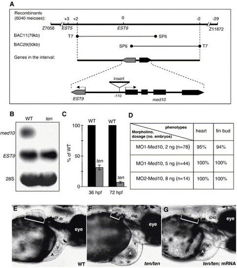
med10 is the gene for tennismatch (ten). (A) Positional cloning of the ten locus. ten was mapped between SSLP markers Z7058 and Z11872 for chromosome 19. The chromosomal walk was initiated from EST9, which has zero recombinants out of 6040 meioses. A physical contig was constructed consisting of two overlapping BACs covering the ten locus, as each end of the contig picks up two recombinants on either side. Two genes were predicted using computational tools after sequencing these two BACs. The genomic structure of med10 is shown at the bottom (GenBank Accession Number AY698065). EST9 is 4 kb away from med10 and is transcribed in the opposite direction. A 1.2 kb sequence (GenBank Accession Number AY698066) was identified to be inserted before the transcriptional start site of med10 in ten mutant zebrafish. (B) Expression of med10 mRNA was abolished in ten mutant embryos. Shown are Northern blots of total RNA extracted from embryos at 42 hpf. (C) Expression of med10 mRNA was quantified by real-time RT–PCR analysis in embryos collected at 36 hpf, when ten mutant embryos start to exhibit phenotypes, and at 72 hpf. Data shown here are the means ± SD from three independent experiments, expressed as a percentage of wild-type transcripts. (D) Injection of morpholino antisense oligonucleotides against med10 phenocopies ten mutant embryos. Injected embryos were evaluated at day 3 based on two visible phenotypes of ten mutants: cardiac malformation and stunted pectoral fin bud growth. Shown are the percentages of embryos that had the corresponding phenotype. (E–G) Injection of med10 mRNA rescues ten mutant phenotypes. Shown are wild-type (E), ten/ten mutant (F), and ten/ten mutant plus med10 mRNA (2 ng) injection (G) at 72 hpf. Anterior is to the right and dorsal is up. The heart is located ventral to the yolk. The brackets indicate the pectoral fin bud. V, ventricle; A, atrium. |
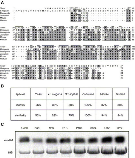
Med10 is a conserved subunit of the transcriptional Mediator complex (MED). (A) Med10 is evolutionarily conserved. The predicted protein sequence for Med10 was compared using ClustalW with Med10 orthologues from several species. (B) Zebrafish Med10 is highly homologous with human and mouse Med10 but exhibits limited homology with Drosophila, C. elegans, and yeast. Shown are percentages of identity and similarity. (C) med10 is expressed throughout zebrafish embryogenesis. Shown is a Northern blot with total RNA extracted from 10 embryos at different stages. Note the decrease of maternal transcripts at the bud stage and gradual increase of zygotic transcripts thereafter. EXPRESSION / LABELING:
PHENOTYPE:
|
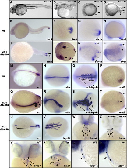
Reduction of Med10 results in specific developmental defects in a dose-dependent manner. (A–D) Reduction of Med10 leads to three classes of defects: (A) Class I embryos, (B) Class II embryos, (C) Class III embryos. The tails in approximately 14% (12/88) of Class III embryos later extend out from the body and clearly exhibit multiple tail buds (D). (E–V) Injection of high dosages of MO1-Med10 (15 ng) leads to altered notochord extension and tail bud formation. In situ hybridization was performed on 30 hpf injected embryos or the age-matched (by counting the somite number) control embryos. Two tail buds can be detected in Class II mutants, as shown by tbx6 staining (I). Approximately four tail buds formed in Class III embryos, as shown by tbx6 (J), cdx1a (K), eve1 (L), ntl (Q,U), and wnt8 (T) staining. The notochord in Class III embryos is indicated by ntl (Q,U) and shh staining (R,S). The somites in Class III embryos are indicated by MyoD staining (S,V). Arrows indicate positions of the multiple tail buds. Shown are dorsal views (F,G,J–O,Q–V) and lateral views (E,H–I,P) with anterior to the left. (W) Injection of low dosages of MO1-Med10 (5 ng) leads to whole-heart expression of bmp4, which can be rescued to the cardiac cushion-restricted expression pattern by co-injection with med10 mRNA (2 ng) (X). (Y-b) ten mutants exhibit cardiac cushion defects. Shown are bmp4 expression in wild-type (Y) and ten mutant (Z) embryos, and notch1b expression in wild-type (a) and ten mutant (b) embryos. Arrows indicate the atrial–ventricular cushion. Shown in (W–Z) are ventral views of 48 hpf embryos. Anterior is up. Shown in (a,b) are lateral views of 48 hpf embryos. Anterior is left and dorsal is up. V, Ventricle. A, Atrium. EXPRESSION / LABELING:
PHENOTYPE:
|

Reduction of Med10 enhances Wnt signaling. (A–H) Reduction of Med10 results in expanded expression of Wnt target genes. MO1-Med10 (5 ng) was injected into embryos at the one-cell stage. Embryos were then fixed at either 30% epiboly (A–D) or 75% epiboly (E–H) and stained using in situ hybridization. Dorsal is right. Animal views are shown in (A–D); lateral views are shown in (E–H). eve1 expression was not affected by reduction in Med10 (A,B), serving as a control. Shown are spr2 expression in MO1-Med10 injected embryos (D,F) and control embryos (C,E); and cdx4 expression in MO1-Med10 injected embryos (H) and control embryos (G). (I–N) Reduction of Med10 leads to a graded expansion of lef1:gfp. Increasing amounts of MO1-Med10 were injected into embryos at the one-cell stage and in situ hybridization using a gfp riboprobe was performed on embryos at 30% (I–K) and 90% (L–N) epiboly. Animal pole views are shown in (I–K); lateral views are shown in (L–N). The region of gfp expression is increased in med10 morphants (J,K,M,N), as compared to control embryos (I,L). (O–Q) Reduction of Med10 enhances Wnt8-induced ectopic tail bud formation. wnt8/gfp mRNA (100 pg each) were injected into single cells at the animal pole of 128-cell stage embryos. Ectopic tail buds were induced in GFP-positive embryos (O), as confirmed by staining for tbx6 (P). Arrows indicate ectopic tail buds (O,P). (Q) Quantification of Wnt8-induced ectopic tail buds. Data presented in (Q) are the mean ± SD from at least three independent experiments. Total embryos scored were 663, 258, and 129 for wnt8/gfp, wnt8/gfp/MO1-Med10 (100 pg/100 pg/150 pg) and wnt8/gfp/med10 mRNA (100 pg each) injections, respectively. EXPRESSION / LABELING:
|
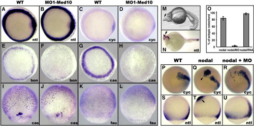
Reduction of Med10 inhibits Nodal signaling. (A–H) Reduction of Med10 suppresses Nodal target-gene expression. MO1-Med10 (15 ng) was injected into embryos at the one-cell stage, and embryos were fixed at 30% epiboly and stained using in situ hybridization. Shown are animal pole views. Shown are nodal (cyclops) and ntl expression in MO1-Med10 injected (B,D) and control (A,C) embryos; and bon and cas expression in MO1-Med10 injected (F,H) and control (E,G) embryos. (I–L) Reduction of Med10 leads to an altered expression pattern of Nodal target genes. Embryos injected with MO1-Med10 (5 ng) at the one-cell stage were fixed at 75% epiboly and subject to in situ hybridization staining. Shown are lateral views with the animal pole up. Mesoendoderm cells, as stained by cas and fau, moved faster toward the animal pole in the med10 morphants (J,L), when compared with controls (I,K). (M–U) Reduction of Med10 suppresses Nodal-induced ectopic notochord formation. Single-cell injection of nodal (cyclops)/gfp mRNA (10 and 100 pg, respectively) at the 128-cell stage induced ectopic notochords in one-day old embryos (M), as confirmed by ntl staining (N). Ectopic ntl expression could be detected as early as 75% epiboly in nodal (cyclops)/gfp injected embryos (T), but not in embryos co-injected with MO1-Med10 (150 pg) (U) and control embryos (S). In addition, note the clustered and scattered expression of ectopic nodal (cyclops) in embryos injected with nodal (cyclops)/gfp and nodal (cyclops)/gfp/MO1-Med10, respectively (Q,R). (O) Quantification of Nodal-induced ectopic notochords. Data presented in (O) are the means ± SD from at least three independent experiments. Total embryos scored were 735, 140, and 183 for nodal (cyclops)/gfp, nodal (cyclops)/gfp/MO1-Med10 (10 pg/100 pg/150 pg), and nodal (cyclops)/gfp/Med10 mRNA (10 pg/100 pg/100 pg) injections, respectively. Arrows indicate ectopic notochord formation (M,N,T), and the arrowhead indicates ectopic nodal (cyclops) expression (Q). EXPRESSION / LABELING:
|
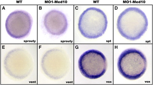
Reduction of Med10 does not affect FGF or BMP signaling. MO1-Med10 (10 ng) was injected into embryos at the one-cell stage. Embryos were fixed at 50% epiboly and stained using in situ hybridization. Animal views are shown. (A–D) Reduction of Med10 has no effect on FGF-regulated gene expression, as manifested by sprouty and spadetail (spt) staining in med10 morphants (B,D) and control embryos (A,C). (E–H) Reduction of Med10 has no effect on the expression of BMP target genes as manifested by in situ hybridization staining for vent and vox (F,H), as compared to uninjected control embryos (E,G). Shown are animal pole views with dorsal to the right. EXPRESSION / LABELING:
|
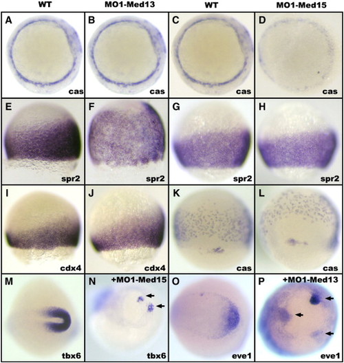
The multiple tail bud phenotype of med10 morphants is recapitulated in med13/med15 double morphants. Injection of MO1-Med13 (10 ng) expands Wnt target-gene expression. Shown are spr2 and cdx4 staining in injected (F,J) and control (E,I) embryos. The expression of cas (A,B) serves as a control. Injection of MO1-Med15 (10 ng) suppresses Nodal target gene cas expression (D), but does not affect the expression of spr2 (H), as compared to control embryos (C,G). Mesoendoderm cells, as detected by staining for cas, are expressed in a region more close to the animal pole in med15 morphants (5 ng of MO1-Med15 injection) (L), as compared to control embryos (K). Shown in (A–D) are animal pole views of 30% epiboly embryos. Shown in (E–L) are lateral views of 75% epiboly embryos. (M–P) Reduction of both Med13 and Med15 (15 ng of MO1-Med13 and 3 ng of MO1-Med15) recapitulates the multiple tail bud phenotype of med10 morphants, as shown by tbx6 (N) and eve1 (P) staining. Shown are dorsal views. Arrowheads indicate the position of the ectopic tail buds. EXPRESSION / LABELING:
PHENOTYPE:
|
Reprinted from Developmental Biology, 303(2), Lin, X., Rinaldo, L., Fazly, A.F., and Xu, X., Depletion of Med10 enhances Wnt and suppresses Nodal signaling during zebrafish embryogenesis, 536-548, Copyright (2007) with permission from Elsevier. Full text @ Dev. Biol.