
Fig. 1
|
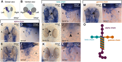
Fig. 1 Dynamic expression of laminin subunit genes during heart morphogenesis. (A,B) Schematic of the position of the heart (blue) in a 30 hpf zebrafish embryo (A; dorsal view) and a 55 hpf zebrafish embryo (B; ventral view). The embryo body is shaded pale grey, eyes are shaded dark grey and the yolk is shaded in yellow. (C-F) mRNA in situ hybridisation expression analysis of laminin alpha chain subunits lama4 (C,D) and lama5 (E,F) in the heart. (G-L) mRNA in situ hybridisation expression analysis of laminin beta subunit chains lamb1a (G,H), lamb1b (I,J) and lamb2 (K,L) in the heart. (M,N) mRNA in situ hybridisation expression analysis of gamma subunit lamc1 in the heart. Arrowheads indicate the position of the heart and the anterior is to the top in all images. (O) Schematic of the heterotrimeric structure of laminin. Scale bars: 50 μm. A, atrium; V, ventricle.