
Fig. 4
|
Fig. 4
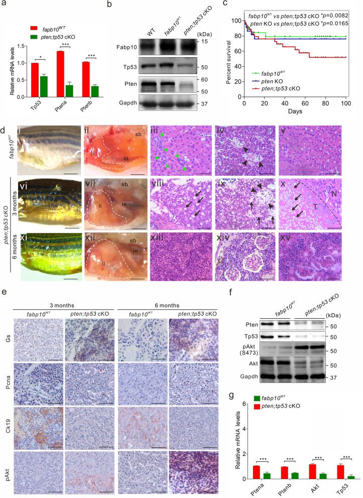
Mutation of tp53 is critical for the progression of hepatocarcinogenesis. a Abundances of Ptena, Ptenb, or Fabp10 mRNA in liver tissues of fabp10WT and pten;tp53 cKO fish (n = 3 per group). b Western blot analysis of Fabp10, Tp53, and Pten in liver tissues of WT, fabp10WT, and pten;tp53 cKO fish (n = 3 per group). c Overall survival rates of WT, fabp10WT, and pten;tp53 cKO fish (n = 200 per group). d Gross morphology of 3-month-old fabp10WT (di) and pten;tp53 cKO fish (dvi) and 6-month-old pten;tp53 cKO fish (dxi). Representative bright field images of the internal abdominal organs, with the liver outlined, in 3-month-old fabp10WT (dii) and pten;tp53 cKO fish (dvii) and 6-month-old pten;tp53 cKO fish (dxii). in, intestine; li, liver tissues, sb, swim bladder. Histological examination of liver tissues from fabp10WT and pten;tp53 cKO fish at 3 and 6 months of age, respectively. Several typical hepatocarcinogenesis phenotypes were observed in 3- and 6-month-old pten;tp53 cKO fish, including abnormal lipid accumulation in hepatocytes (black arrows, dviii), bile duct disappearance (green arrowheads, dviii-x), vascular disorder (black arrowheads, d9), variation in nuclear/cellular sizes (pleomorphism; dvii, viii), and tumour cell invasion into blood vessels (dxiii), the pancreas (dxiv), and the kidney (dxv). Scale bars, 100 μm. e Immunohistochemical staining was performed to examine the expression of Gs, Pcna, Ck19, and pAkt in liver tissues from 3- and 6-month-old fabp10WT and pten;tp53 cKO fish. Scale bars, 100 μm. f Western blot analyses were performed to confirm the expression of Pten, Tp53, pAkt, and Akt in liver tissues from 3-month-old fabp10WT (lanes 1–2) and pten;tp53 cKO fish (lanes 3–4). g Quantitative analysis of the protein expression ratio of pAkt and Akt in liver tissues from 3-month-old fabp10WT and pten;tp53 cKO fish. The data are shown as the mean ± SEM values. *p < 0.05, ***p < 0.001