
Fig. 5
|
Fig. 5
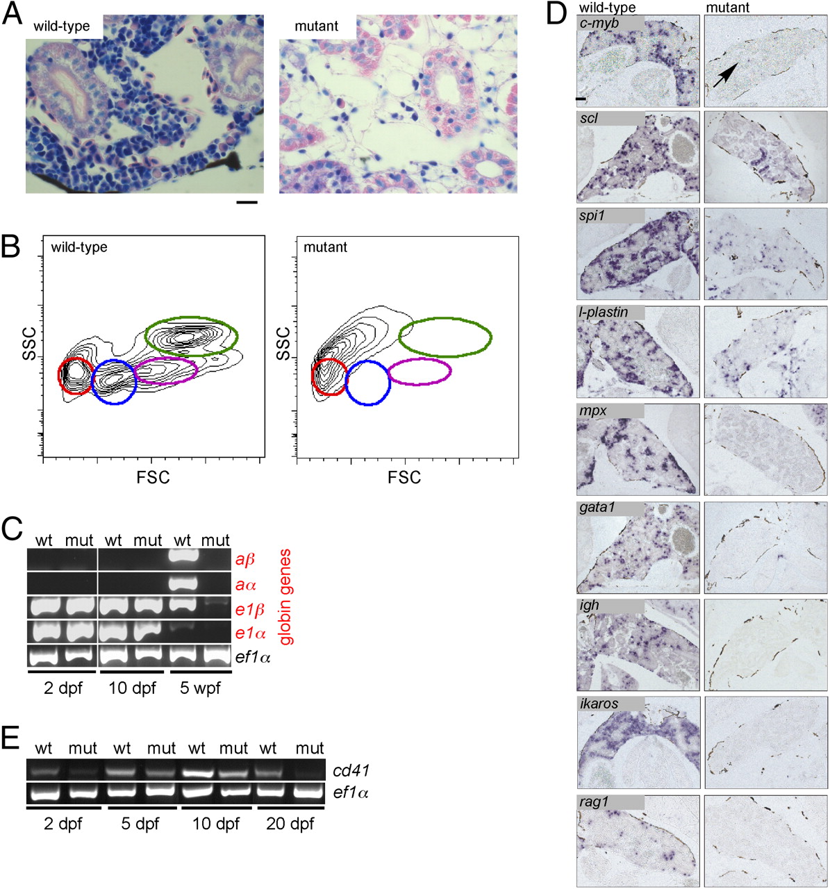
Failure of adult hematopoiesis in c-myb mutants. (A) Histological sections of the head kidney at 7 wk of age (Giemsa staining). Note the lack of hematopoietic cells in the mutant tissue. (Scale bar: 10 μm.) (B) Flow cytometric analysis of whole kidney marrow (6 wk of age) according to side scatter (SSC) and forward scatter (FSC) characteristics. Circles denote the positions of various cell types detectable in wild-type fish: red, erythrocytes; blue, lymphocytes and thrombocytes; magenta, precursors; green, myelomonocytes. The residual cells obtained from disintegrated mutant kidney tissue lack these characteristic features. (C) Lack of adult hemoglobin gene expression in c-myb mutants. RT-PCR was performed at the indicated time points for adult (prefix a) and embryonic (prefix e) globin genes; ef1α serves as a control for cDNA integrity and as a standard. (D) RNA in situ hybridization of head kidney sections was performed with the indicated probes. The signals seen with spi1 and l-plastin presumably originate from long-lived embryonic macrophages; a single c-myb positive cell is indicated (arrow). In mutant tissue, no signals are observed for mpx, gata1, ikaros, rag1, and igh. (Scale bar: 100 μm.) (E) Diminishing expression of cd41 in mutant embryos and larvae. RT-PCR was performed at the indicated time points; ef1α serves as a control for cDNA integrity and as a standard.