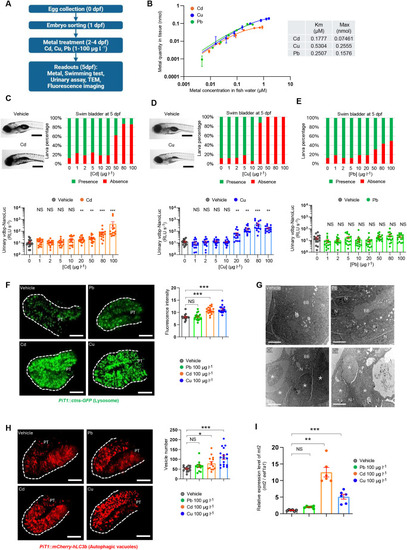
vdbp-NanoLuc reporter as biosensor for detecting metal nephrotoxicity. (A) Experimental protocol of metal toxicity assessment using transgenic ½vdbp-NanoLuc zebrafish larvae. (B) Levels of cadmium (Cd), copper (Cu) and lead (Pb) in larval lysates were analyzed by using inductively coupled plasma mass spectrometry (ICP-MS) after 3 days of larvae incubation with metals. The Michaelis–Menten constant (Km) and maximal quantity in larvae (Max) are calculated from the measurement by using the molar concentration. The Km of Pb is very similar to that of Cd, which is much lower than that of Cu, showing the lower affinity for uptake of Cu by larvae. (C) Morphological examination of 5 dpf larvae treated with vehicle or Cd (top left). Scale bars: 0.5 mm. Semi-quantitative scoring of swim bladder defects and urinary analysis for 5 dpf larvae treated from 2 dpf with Cd at concentrations from 1 to 100 µg l−1. Swim bladder inflation is impaired by Cd treatment at 50 µg l−1 (top right). Excessive urinary loss of vdbp-NanoLuc using luminometry was observed at 20 µg l−1 (bottom). n=16. (D) Morphological examination of 5 dpf larvae treated with vehicle or Cu (top left). Scale bars: 0.5 mm. Semi-quantitative scoring of swim bladder defects and urinary analysis for 5 dpf larvae treated with Cu at concentrations from 1 to 100 µg l−1. Failure to inflate the swim bladder (top right) and excessive urinary loss of vdbp-NanoLuc (bottom) were observed in larvae treated with Cu from the concentration of 20 µg l−1. n=16. (E) Semi-quantitative scoring of swim bladder defects and urinary analysis for 5 dpf larvae treated with Pb at concentration from 1 to 100 µg l−1. No low-molecular-weight (LMW) proteinuria was seen in any Pb-treated group. n=16. (F) Microscopic examination of 5 dpf Tg(PiT1::ctns-EGFP) zebrafish larvae expressing lysosomal marker Ctns-EGFP in the PT of larvae treated with vehicle, Pb, Cd or Cu by using multiphoton fluorescence microscopy (left). Accumulation of Ctns-EGFP-positive vesicles in the PT of larvae treated with 100 µg l−1 Cd or Cu (right). PT, proximal tubule. n=15 (vehicle), n=18 (Pb), n=17 (Cd), n=16 (Cu). Scale bars: 30 µm. (G) The ultrastructure of PT in 5 dpf larvae treated with vehicle, Pb, Cd, and Cu was analyzed by transmission electron microscopy. Accumulation of large electron-dense vesicles corresponding to lysosomes is observed in epithelial cells of larvae treated with 100 µg l−1 Cd or Cu. n=5. Asterisks indicate large electron dense vesicles. BB, brush border; N, nucleus. Scale bars: 5 µm. (H) Microscopic examination of 5 dpf Tg(PiT1::mCherry-hLC3b) zebrafish larvae expressing autophagy marker, mCherry-hLC3b, in PT of larvae treated with vehicle, Pb, Cd or Cu using multiphoton fluorescence microscopy (left). Accumulation of mCherry-hLC3b-positive autophagic vacuoles is observed in PT of larvae treated with 100 µg l−1 Cd or Cu (right). n=14 (vehicle), n=13 (Pb), n=14 (Cd), n=18 (Cu); Scale bars: 30 µm. (I) Analysis of metallothionein mt2 mRNA expression levels in metal-treated larvae by quantitative PCR. Total mRNA was extracted from larvae treated with vehicle or 100 µg l−1 Pb, Cd or Cu. After reverse transcription, the cDNA was analyzed by quantitative PCR to assess mt2 mRNA expression levels. n=6. Plotted data represent mean±s.e.m. Nonparametric Mann–Whitney test, *P<0.05, **P<0.01, ***P<0.001. NS, non-significant.
|
