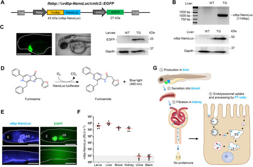
Establishment of the ½vdbp-NanoLuc zebrafish reporter system. (A) Construction of plasmid vector lfabp::½vdbp-NanoLuc containing the transgenesis marker cmlc2::EGFP and Tol2 transposon elements. The N-terminal 207 amino acids of zebrafish VDBP (½vdbp) are fused with NanoLuc luciferase, which is predicted to have a molecular weight of 43 kDa. The liver fatty acid binding protein (lfabp) promoter drives the expression of vdbp-NanoLuc in the liver, and the cardiac myosin light chain 2 (cmlc2; also known as myl7) promoter controls the expression of EGFP in the heart. Tol2, transposable elements facilitating the integration of plasmid DNA into the embryonic genome. (B) Analysis of ½vdbp-NanoLuc cDNA (1144 bp) after amplification by polymerase chain reaction (top) and immunoblotting analysis of vdbp-NanoLuc (bottom) in liver lysate. n=3 replicates. TG, transgenic zebrafish; WT, wild type. (C) Observation of EGFP fluorescence in the heart of transgenic larvae visualized at 2 days post-fertilization (dpf) by light-sheet microcopy (left) and detection of EGFP (27 kDa) by immunoblotting in larval lysate from WT and transgenic ½vdbp-NanoLuc larvae (right). n=3 replicates; one sample is a pool of five larvae. Dotted lines outline the larva head/body. Scale bar: 0.5 mm. (D) The oxidation of furimazine by NanoLuc luciferase in the presence of O2 produces furimamide, CO2 and blue light with a wavelength of 460 nm. (E) Observation of ½vdbp-NanoLuc larva at 5 dpf by bioluminescence microscopy using an Olympus LV200. With incubation of the furimazine substrate, bioluminescent light from vdbp-NanoLuc (blue) is seen at least in the liver and vessels, with EGFP fluorescence (green) visible in the heart. n=3. Dotted lines outline the larva head/body, liver and heart. E, eye; H, heart; L, liver; SB, swim bladder. Scale bars: 0.5 mm. (F) Quantification of vdbp-NanoLuc luciferase activity in transgenic zebrafish by luminometry: whole larval lysates (lane 1), isolated liver (lane 2), blood (lane 3), isolated kidney (lane 4), urine collected from 5 dpf larvae (lane 5), blank (E3 medium, lane 6); n=6. (G) Schematic illustration of the fate of recombinant protein vdbp-NanoLuc under normal physiological conditions: vdbp-NanoLuc is produced in liver ①, secreted into the bloodstream ②, passed through the glomerulus filtration in kidney ③, reabsorbed and processed by proximal tubule epithelial cells ④. EE, early endosome; LE, late endosome; L, lysosome; PT, proximal tubule.
|
