
Fig. 5
- ID
- ZDB-FIG-250505-73
- Publication
- Yang et al., 2025 - The Serotonergic Dorsal Raphe Promotes Emergence from Propofol Anesthesia in Zebrafish
- Other Figures
- All Figure Page
- Back to All Figure Page
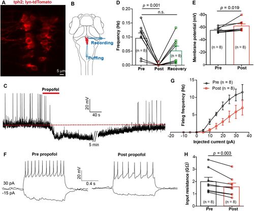
In vivo whole-cell recording revealed that propofol decreased the excitability of DRN5-HT. A, The expression of Ki(tph2:GAL4FF,cmlc2:EGFP);Tg(UAS: lyn-tdTomato) mutant line at dorsal raphe. B, Schematic diagram showing whole-cell recording of DRN5-HT neurons and simultaneous local puffing of propofol onto DRN5-HT. C, Spontaneous spike firing of DRN5-HT neuron before and after puffing of propofol. The red dashed line indicates the mean resting membrane potential before propofol treatment. D,E, Summary of data showing the effect of propofol treatment on the spontaneous spike frequency (D, pre vs post, 0.10 ± 0.02 Hz vs 0.00 ± 0.00 Hz; p = 0.001; pre vs recovery, 0.10 ± 0.02 Hz vs 0.05 ± 0.02 Hz; p > 0.05; paired Student's t test) and resting membrane potential (E, pre vs post, −55.1 ± 1.5 mV vs −62.0 ± 3.1 mV; p = 0.019; paired Student's t test) of DRN5-HT neurons. Pre, before puffing; post, immediately after puffing; recovery, 10 min after puffing. F, Representative traces showing voltage responses of DRN5-HT evoked by current injections before (left) and immediately after (right) puffing of propofol. G, Summary of data showing the effect of propofol on current injection-evoked spike firing of DRN5-HT neurons. p = 0.01, Kolmogorov–Smirnov test. H, Summary of data showing the effect of propofol on the membrane resistance of DRN5-HT neurons. Pre versus post, 2.0 ± 0.3 versus 1.6 ± 0.3 GΩ; p = 0.019; paired Student's t test. Numbers of cells examined are in parentheses. |