
Fig. 3
- ID
- ZDB-FIG-250505-71
- Publication
- Yang et al., 2025 - The Serotonergic Dorsal Raphe Promotes Emergence from Propofol Anesthesia in Zebrafish
- Other Figures
- All Figure Page
- Back to All Figure Page
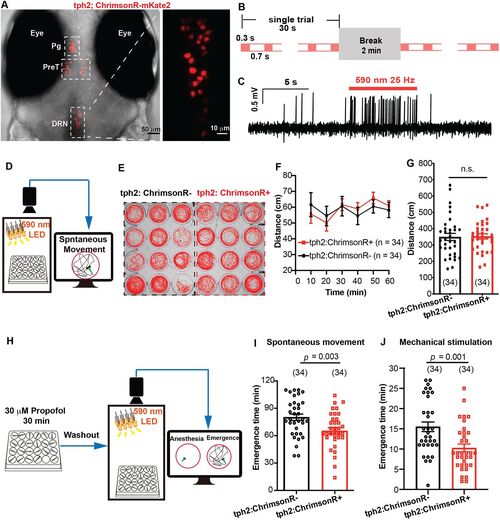
Optogenetic activation of tph2+ neurons promoted emergence from propofol anesthesia. A, The expression of Ki(tph2: GAL4FF,cmlc2:EGFP);Tg(UAS: ChrimsonR-mKate2) mutant line. Pg, pineal gland; PreT, pretectum; DRN, dorsal raphe nucleus. B, Optogenetic stimulation paradigm during the whole process of emergence. C, Confirmation of ChrimsonR activation by in vivo cell-attached recording. D, The diagram of behavior experiment detecting the influence of field optogenetic activation on locomotor activity. E, The spontaneous movement trajectory under optogenetic stimulation during 10 min. F,G, Distance moved under optogenetic stimulation during 1 h and the statistical analysis (tph2, ChrimsonR−, vs tph2, ChrimsonR+; 349.8 ± 21.7 mm vs 353.0 ± 15.8 mm; p > 0.05; unpaired Student's t test). H, The diagram of behavioral experiment detecting the influence of field optogenetic activation on the emergence time from propofol. I,J, Optogenetic stimulation significantly promoted the emergence from propofol anesthesia detected by recovery of spontaneous movement and recovery to mechanical stimulation (spontaneous movement, tph2, ChrimsonR−, vs tph2, ChrimsonR+; 80.2 ± 3.5 min vs 65.3 ± 3.3 min; p = 0.003; mechanical stimulation, tph2, ChrimsonR−, 15.5 ± 1.2 min; tph2, ChrimsonR+, 10.3 ± 1.0 min; p = 0.001; unpaired Student's t test). The sample size for each group is indicated in parentheses. |