
Fig. 5
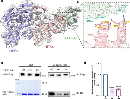
The interaction between BLOC-3 and RAB9A.a Low-resolution cryoEM envelope of BLOC-3-Rab9A Q66L with docked AlphaFold3 model. b Close-up view of the HPS4-RAB9A binding interface, with critical residues for RAB9A binding shown in sticks. Residues that are mutated in (d) are indicated with a yellow box. c Flag-tagged HPS4 WT and mutants were expressed and purified using HEK293T cells, then incubated with purified His-tagged mouse Rab9A Q66L mutant and subjected to Flag pull-down. HPS4 and Rab9A were detected by antibodies against Flag and His, respectively. d Quantification of the results presented in (c). Statistical differences were determined by One-way ANOVA. Error bars represent the standard deviation of three independent biological repeats. n = 3. |