|
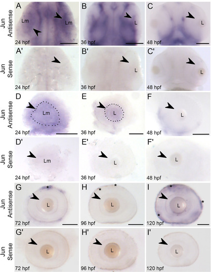
Jun expression in the developing retina is downregulated by 48 hpf. Whole mount in situ hybridization of jun expression in Tg(isl2b:GFP) embryos and larvae from 24 hpf – 120 hpf. (A-C) jun expression in the anterior region of the embryo at 24, 36, and 48 hpf, dorsal views. A. Arrowhead indicates jun expression in the optic lobe. B. Arrowhead indicates jun mRNA staining in the retinal progenitor cell layer. C.jun expression is absent in the ganglion cell layer, arrowhead. (A’-C’) jun sense controls in the anterior region. (D-I’) jun expression and sense controls in lateral views of dissected eyes from 24 hpf - 120 hpf. D.jun expression is present throughout the optic cup. A dotted line outlines the lens mass (Lm) and arrowhead indicates the most concentrated jun expression surrounding the lens mass. E. The retinal progenitor cell layer displays jun expression, outlined. This expression, arrowhead, cups the lens. (F-I) jun expression is undetectable in the ganglion cell layer at 48 hpf- 120 hpf. Arrowheads indicate the ganglion cell layer of the now formed retina. Asterisks indicate the outer pigmented epithelial layer. (A-C) Anterior is toward the top, A-B, dorsal view, C, ventral view. A,D, n = 44; A’,D’ n = 6; B,E, n = 47; B’,E’, n = 5; C,F, n = 34; C’,F’, n = 4; G, n = 23; G’, n = 8; H, n = 26; H’, n = 4; I, n = 25; I’, n = 7. Lm = lens mass; L = lens. Scale bars = 100 µm.
|