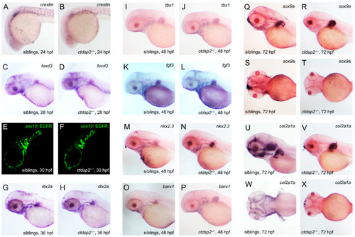
The impact of ctdsp2 knockout on pharyngeal pouches, neural crest cells (NCCs), and pharyngeal cartilage development in zebrafish embryos: (A–D) ISH results with crestin and foxd3 probes, markers for NCCs, at 24 h post-fertilization (hpf) and 28 hpf. The staining patterns show no noticeable differences between mutant embryos and their control siblings. (E,F) Fluorescence imaging of sox10-labeled NCCs in the pharyngeal arch region. Green fluorescence signals indicating NCCs are similar in both ctdsp2−/− embryos and siblings. (G,H) Expression of dlx2a at 30 hpf appears similar in both ctdsp2−/− and control embryos. (I–N) Expression of tbx1, fgf3, and nkx2.3 at 48 hpf shows no significant differences in the segmentation and number of pharyngeal pouches between ctdsp2−/− embryos and siblings. (O,P) ISH with the barx1 probe at 48 hpf. No significant variation was observed between mutant and control embryos. (Q–X) ISH with sox9a and col2a1a probes at 72 hpf; sox9a expression is notably reduced in mutants, and the expression of col2a1a, a marker for cartilage, is absent in the hypopharyngeal arches of the mutant embryos compared to controls.
|
