FIGURE

Figure 7

ID
ZDB-FIG-250215-59
Publication
Xia et al., 2025 - ctdsp2 Knockout Induces Zebrafish Craniofacial Dysplasia via p53 Signaling Activation
Other Figures
All Figure Page
Back to All Figure Page
Figure 7

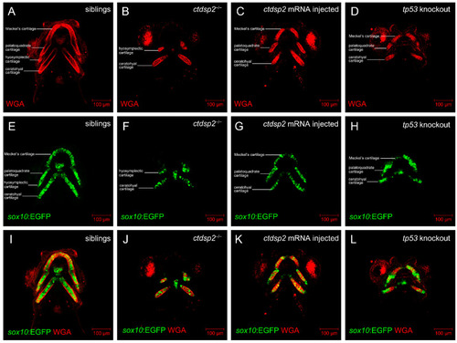
Comparative analysis of chondrocyte morphology in (A,E,I) control siblings, (B,F,J) ctdsp2−/− mutants, (C,G,K) ctdsp2−/− embryos injected with ctdsp2 mRNA, and (D,H,L) tp53-knockout ctdsp2−/− embryos. The craniofacial cartilage in control siblings demonstrates a consistent, elongated, and slender chondrocyte structure, forming a “stack of pennies” organization. In ctdsp2−/− embryos, chondrocytes within the cartilage are noticeably smaller, and the overall cartilage structures are markedly deformed. Injection of ctdsp2 mRNA or knockout of tp53 in mutants partially rescued chondrocyte morphology.

Expression Data

Expression Detail
Antibody Labeling
Phenotype Data

Phenotype Detail
Acknowledgments
This image is the copyrighted work of the attributed author or publisher, and ZFIN has permission only to display this image to its users. Additional permissions should be obtained from the applicable author or publisher of the image. Full text @ Int. J. Mol. Sci.