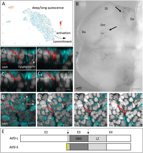
atf3 is expressed in a subset of NSCs in the adult pallium. (A) atf3pos cells (orange dots, UMI values color-coded) on the scRNAseq UMAP of adult quiescent NSCs (blue dots). NSCs closest to activation and/or neurogenesis commitment and NSCs in deep/long quiescence are indicated (Morizet et al., 2024). (B) Expression of atf3 in whole-mount ISH in the adult pallium (dorsal view, anterior left) with the atf3-L probe shown in E. Da, Dl, Dm, Dp, anterior, lateral, median and posterior pallial domains. Arrows point to atf3pos cells in Dm and Dp. (C1-C4) Optical cross-sections of a Tg(gfap:eGFP) pallium stained in whole-mount for GFP (cyan, IHC), atf3 transcripts (Fast Red) and DAPI (ventricular surface up). Sections are ordered from C1 to C4 along the anteroposterior axis. Red arrows indicate an atf3pos, GFPpos cell (asterisk indicates the nucleus). (D1-D4) Optical horizontal sections of the pallium shown in C. D1 to D4 show superficial to deeper locations in sequence. Red arrows indicate an atf3pos, GFPpos cell. (E) Atf3 protein isoforms predicted from adult pallial transcripts. DBD, DNA-binding domain; E2-E4, coding exons: L, long; LZ, leucine zipper; S, short. Arrows indicate splice junctions for the L form. Yellow box represents the N terminus of the S form (alternative splicing). Scale bars: 100 µm (B); 5 µm (C1-D4).
|
